
The Radiology Partners (RP) Cardiothoracic Imaging National Subspecialty Division (NSD) presents our newest Rad to Rad Learning case.
Peer Learning Opportunity

Although infrequent, intra-cardiac thrombi or masses have significant ramifications and can result in pulmonary or systemic embolization.
Incidental Intra-Cardiac Lesions
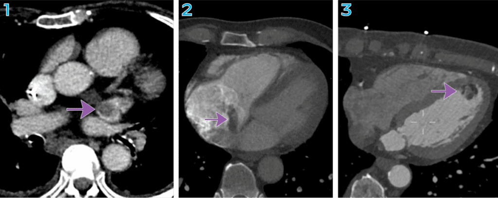
All three images are from CT abdomen/pelvis exams.
1. Left atrial appendage thrombus
2. Right artial mass.
3. Left ventricular mass.
Shared to improve patient safety and healthcare delivery in the provision of radiology services. The circumstances and facts are changed, altered, or deidentified to preserve confidentiality. Privileges have not been waived.
Practical Insights
-
-
Though motion artifacts and contrast flow artifacts can make the heart chambers difficult to assess, include them in your search patterns of non-cardiac examinations.
-
Think beyond heart size and coronary calcifications when reviewing the heart.
The Cardiothoracic Imaging National Subspecialty Division (NSD) is part of RP’s Clinical Value Team, which works to elevate patient care and enhance value through innovation, collaboration and education. To advance this goal, our radiologists and advanced practice providers are committed to sharing peer learning as valuable reminders and insights about what we encounter in our day-to-day practice. Check back here and on X, LinkedIn and Instagram to see these common cases and our findings.
Visit the Clinical Resources page for more cases and to see what we’ve developed to enhance best practice recommendations, elevate image quality and patient care and update current standards throughout RP’s network of practices, all to deliver excellent radiology services to patients, referring clinicians and client partners.

Radiology Partners, through its owned and affiliated practices, is a leading physician-led and physician-owned technology-enabled radiology practice in the U.S. For the latest news from RP, follow us on X, LinkedIn, Instagram, YouTube and the blog.