Dr. Vivek Bansal
In May, we recognize National Stroke Awareness Month.
Last year, we discussed the most common type of stroke with Dr. Vivek Bansal, National Subspecialty Lead for Neuroradiology at Radiology Partners. This National Stroke Awareness Month, he shares information for radiologists on another type, the hemorrhagic stroke, and about the latest technology used to detect them.
Q: Tell us more about the hemorrhagic stroke.
A lot of the coverage of stroke focuses on ischemic strokes, which make up 90% of strokes. The other 10% of strokes are hemorrhagic strokes. While less common, hemorrhagic strokes result in a disproportionate number of stroke-related deaths and can cause severe morbidity.
Hemorrhagic strokes can be broken down into two categories: intracerebral hemorrhage resulting in blood within the brain parenchyma, or subarachnoid hemorrhage with blood surrounding the brain.
Q: What causes this type of stroke?
The most common cause of hemorrhagic stroke is hypertension. In addition, intracranial aneurysms and arteriovenous malformations (AVMs) can also result in bleeding due to weakened blood vessel walls and altered flow dynamics.
Q: How common are aneurysms and AVMs?
In the United States, approximately 1 in 50 people will have an aneurysm. Among those with an aneurysm, 10-20% will have more than one. On average in the United States, there is an aneurysm rupture every 18 minutes, and once ruptured, there is a 40-50% fatality rate. Of the patients who survive the initial event, 50% will have delayed mortality and the remainder often have significant brain deficits.
AVMs occur in less than 1% of the population, but more than 50% of people with an AVM will experience an intracranial hemorrhage. The average age of rupture for an AVM is 17 years old, and the mortality rate for AVMs that hemorrhage is 10-15% with morbidity in 30-50%.
Q: How do radiologists detect and treat this issue?
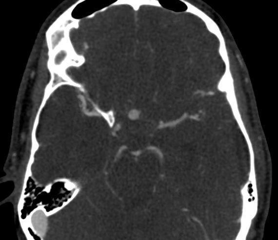
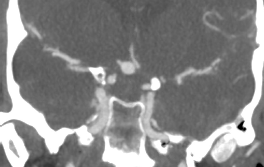
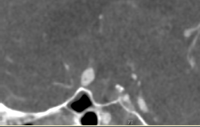
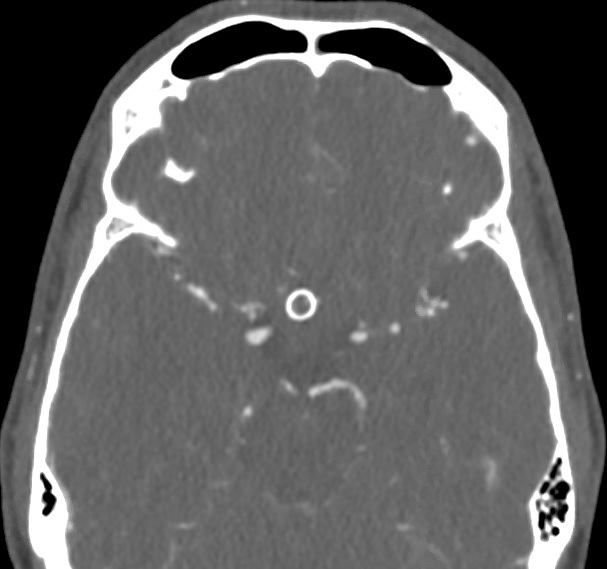
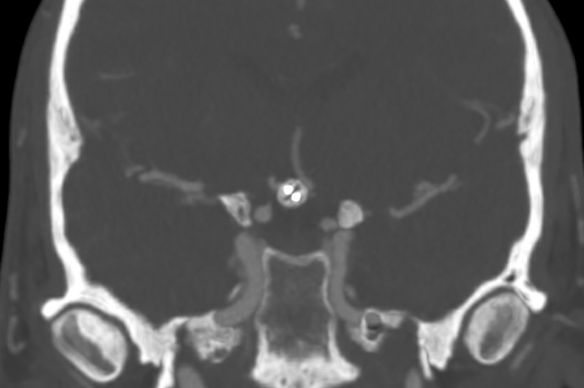
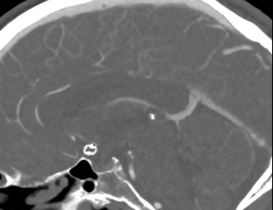
Typically, most brain aneurysms are treated with endovascular coiling and occasionally with neurosurgical clipping. A newer device that is available for endovascular treatment of aneurysms is the woven endobridge. Radiologists should be familiar with the appearance of this device on CT (see figure below). The treatment of arteriovenous malformations is still predominantly through liquid embolic material.
Top row: Axial, coronal and sagittal images of an unruptured anterior communicating artery aneurysm. Bottom row: Axial, coronal and sagittal images after placement of a woven endobridge device. Photo credits: Dr. Benjamin Ludwig at MBB Radiology.
In addition, there are recent advancements in aneurysm detection utilizing artificial intelligence. Aidoc now has Food and Drug Administration approval for their tool to aid radiologists with diagnosis of brain aneurysms on CTA images. With earlier detection, more patients can receive treatment before suffering hemorrhage from rupture.
Dr. Vivek Bansal is a practicing neuroradiologist and the National Subspecialty Lead for Neuroradiology at Radiology Partners, a leading physician-led and physician-owned radiology practice in the U.S. Learn more about our mission, values and practice principles at RadPartners.com. For the latest news from RP, follow along on the RP blog and on Twitter, LinkedIn, Instagram and YouTube.

