
The Radiology Partners (RP) Pediatric Radiology National Subspecialty Division (NSD) presents our newest Rad to Rad Learning case.
Peer Learning Opportunity

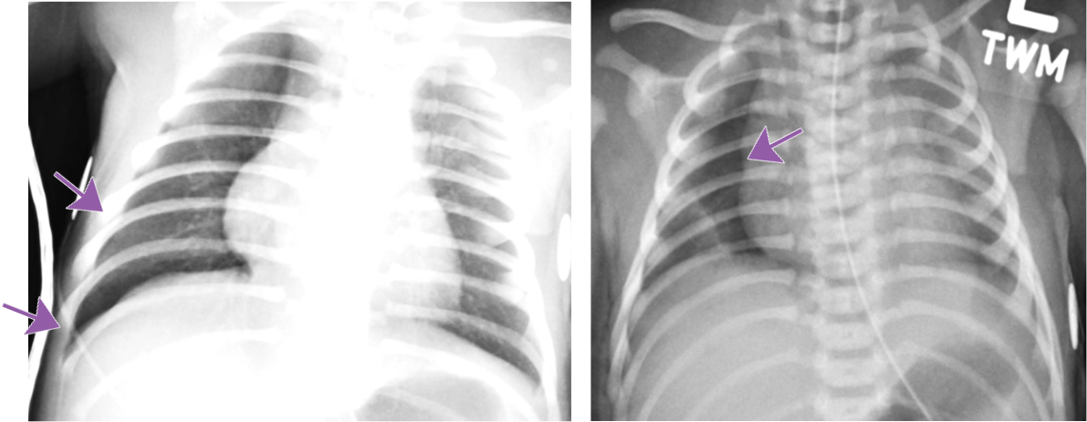
This condition is associated with high morbidity but is difficult to see on supine neonates.
Anterior Pneumothorax
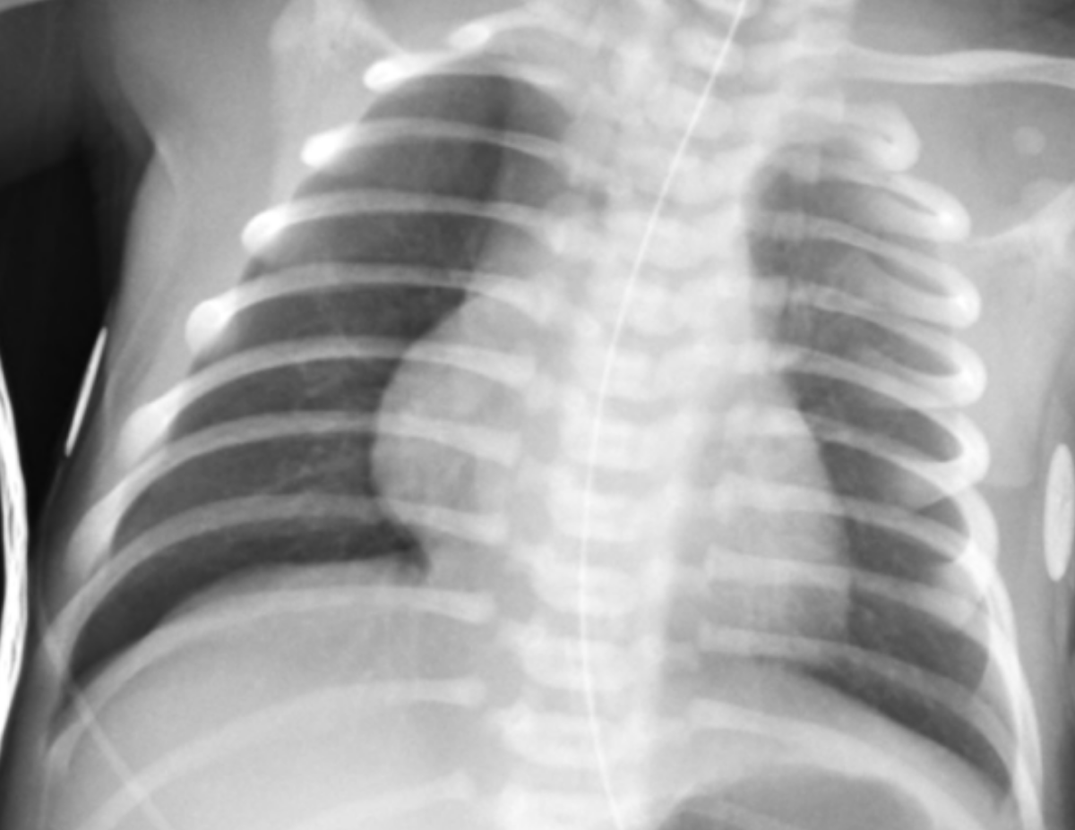
Hallmarks: Deep sulcus sign, no lung marking at the edge of right lung, increased sharpness of the cardiomediastinal border, more prominent on expiration.
Shared to improve patient safety and healthcare delivery in the provision of radiology services. The circumstances and facts are changed, altered, or deidentified to preserve confidentiality. Privileges have not been waived.
Practical Insights
-
-
May be bilateral.
-
Can occur in both term and preterm babies.
-
Compare lung lucency between both sides.
-
Decubitus radiograph can be a helpful tool for confirmation.
-
Takeaway: Even a suspected pneumothorax is a critical result and should be called.
The Pediatric Radiology National Subspecialty Division (NSD) is part of RP’s Clinical Value Team, which works to elevate patient care and enhance value through innovation, collaboration and education. To advance this goal, our radiologists and advanced practice providers are committed to sharing peer learning as valuable reminders and insights about what we encounter in our day-to-day practice. Check back here and on X, LinkedIn and Instagram to see these common cases and our findings.
Visit the Clinical Resources page for more cases and to see what we’ve developed to enhance best practice recommendations, elevate image quality and patient care and update current standards throughout RP’s network of practices, all to deliver excellent radiology services to patients, referring clinicians and client partners.

Radiology Partners, through its owned and affiliated practices, is a leading physician-led and physician-owned technology-enabled radiology practice in the U.S. For the latest news from RP, follow us on X, LinkedIn, Instagram, YouTube and the blog.