Rad to Rad Learning: Three-Phase Bone Scan: Prosthesis Evaluation

The Radiology Partners (RP) NMMI Radiology National Subspecialty Division (NSD) presents our newest Rad to Rad Learning case. 


Peer Learning Opportunitymegaphone icon

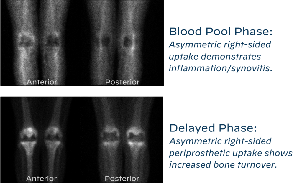
Diagnosis of prosthetic loosening (septic or aseptic) can be achieved with three-phase bone scan using proper technique and specific scintigraphic findings.

63/yo F with right knee pain, bilateral TKAs in 2010
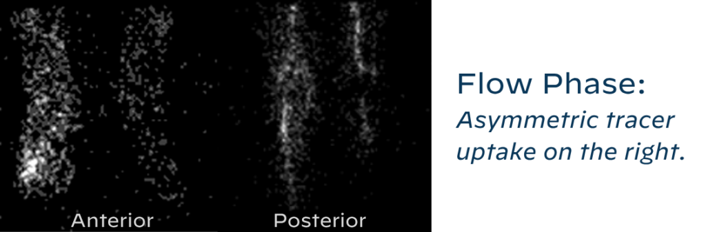
Flow Phase: Asymmetric tracer uptake on the right.
Shared to improve patient safety and healthcare delivery in the provision of radiology services. The circumstances and facts are changed, altered, or deidentified to preserve confidentiality. Privileges have not been waived.


Practical Insights duplicate icon

    • Time since surgery is important history. Patients can have reactive uptake on all three phases up to 1-2 years since surgery.

    • Cemented prostheses usually revert to baseline uptake after one year. Non-cemented prostheses can take up to two years.

    • Laterality and site of pain are also essential history that increase the diagnostic yield.


Bell iconTakeaway: Abnormal uptake in all three phases likely indicates septic loosening.

The The NMMI Radiology National Subspecialty Division (NSD) is part of RP’s Clinical Value Team, whichworks to elevate patient care and enhance value through innovation, collaboration and education. To advance this goal, our radiologists and advanced practice providers are committed to sharing peer learning as valuable reminders and insights about what we encounter in our day-to-day practiceCheck back here and on XLinkedIn and Instagram to see these common cases and our findings.

Visit the Clinical Resources page for more cases and to see what we’ve developed to enhance best practice recommendations, elevate image quality and patient care and update current standards throughout RP’s network of practices, all to deliver excellent radiology services to patients, referring clinicians and client partners.
Radiology Partners, through its owned and affiliated practices, is a leading physician-led and physician-owned technology-enabled radiology practice in the U.S. For the latest news from RP, follow us on XLinkedInInstagramYouTube and the blog.


Radiology Partners Appoints Former Gov. Larry Hogan to Board of Directors

Former Maryland governor brings public health, healthcare innovation and executive leadership expertise to esteemed Board

(NASHVILLETenn.) – May 1, 2026  

(RP), the leading technology-enabled radiology practice in the U.S. through its affiliated practices and Mosaic Clinical Technologies™ (Mosaic), a wholly-owned subsidiary of RP, announced today that former Gov. Larry Hogan has joined its Board of Directors. A nationally recognized leader and cancer survivor, Gov. Hogan brings his deep expertise in public health, visionary leadership and drive for clinical innovation to support RP’s physician-led mission to transform radiology and advance patient-centered care.

“Gov. Hogan brings a unique combination of executive leadership, healthcare policy perspective and personal experience that aligns deeply with our mission,” said Rich Whitney, RP board chair and CEO. “He understands the importance of timely diagnosis, clinical innovation and coordinated care, and he brings insights that will be incredibly valuable as RP continues working to drive clinical, technology and AI innovation to improve patient outcomes.”

Hogan served as the 62nd governor of Maryland from 2015 to 2023 and as chairman of the National Governors Association from 2019 to 2020. Shortly after taking office, he was diagnosed with an aggressive form of non-Hodgkin’s lymphoma and continued to lead the state while undergoing treatment ­­– an experience that shaped his advocacy for early detection, research and innovation in cancer care.

During his tenure, Hogan advanced healthcare and life sciences innovation, including the launch of Maryland’s $216 million Cancer Moonshot initiative to expand cancer detection, treatment, research capacity and workforce development. His administration also prioritized healthcare affordability and access and guided the state through the pandemic, coordinating cross-sector efforts to expand testing, vaccination and the effective delivery of care.

“I’m honored to join the board of Radiology Partners in support of its mission to transform radiology,” said Hogan. “I know firsthand the importance of timely, accurate diagnosis for patients and families alike. As governor, and as chair of the National Governors Association during COVID-19, I saw the essential role physicians and clinical innovation play in strengthening care delivery and improving outcomes. I look forward to working closely with the Radiology Partners team and taking part in the exciting transformative innovation that is underway.”

Gov. Hogan’s perspective, informed by an accomplished public service career and personal healthcare experiences, will support the long-term focus held by RP and Mosaic on physician-led excellence, clinical and technology innovation and high-quality radiology services at scale.

About Radiology Partners

Radiology Partners, through its affiliated practices, is the leading technology-enabled radiology practice in the U.S., serving more than 3,400 hospitals and other healthcare facilities with high quality radiology, technology and artificial intelligence solutions. As a physician-led and physician-owned practice, our mission is to transform radiology by innovating across clinical value, technology, service and economics, while elevating the role of radiology and radiologists in healthcare. Using a proven healthcare services model, Radiology Partners provides consistent, high-quality care to patients, while delivering enhanced value to the hospitals, clinics, imaging centers and referring physicians we serve. Learn more at radpartners.com and connect with us on LinkedIn, X, Instagram and YouTube. 

Media Contacts
Sheila Biggs
sbiggs@jarrardinc.com


Rad to Rad Learning: Pott’s Puffy Tumor

The Radiology Partners (RP) Neuroradiology National Subspecialty Division (NSD) presents our newest Rad to Rad Learning case. 


Peer Learning Opportunitymegaphone icon

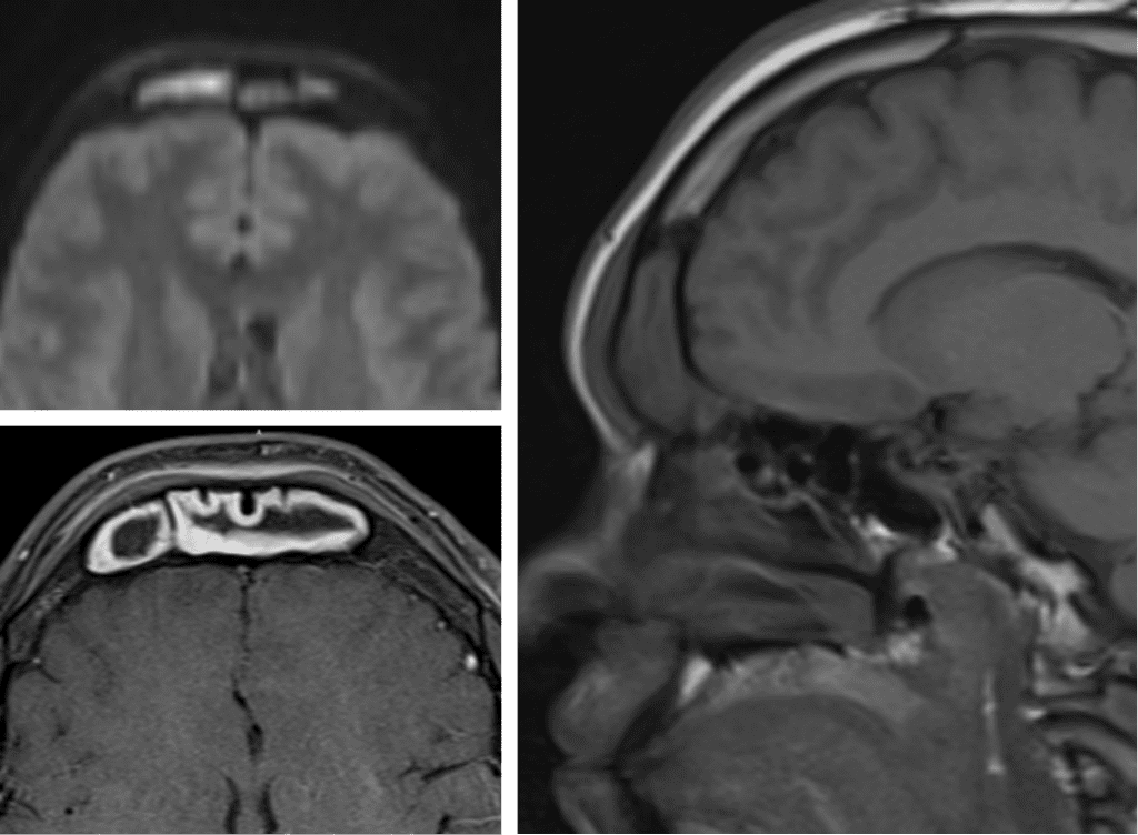
The possibility of intracranial extension of sinus infection results in high morbidity.  

Pott’s Puffy Tumor 

Subperiosteal abscess related to extension of acute frontal sinusitis through the calvarium.

Shared to improve patient safety and healthcare delivery in the provision of radiology services. The circumstances and facts are changed, altered, or deidentified to preserve confidentiality. Privileges have not been waived.

Practical Insights duplicate icon

    • Factors that increase the risk of developing Pott’s include trauma, intranasal cocaine use, methamphetamine use, and craniotomy.

    • Commonly associated intracranial extension needs to be evaluated with an MRI.

    • The overlying calvarium can remain intact.

    • DWI sequence is key for abscess evaluation.

    • More common in adolescents.


Bell iconTakeaway: Prompt identification is required to avoid significant neurologic complications. 

The Neuroradiology National Subspecialty Division (NSD) is part of RP’s Clinical Value Team, whichworks to elevate patient care and enhance value through innovation, collaboration and education. To advance this goal, our radiologists and advanced practice providers are committed to sharing peer learning as valuable reminders and insights about what we encounter in our day-to-day practiceCheck back here and on XLinkedIn and Instagram to see these common cases and our findings.

Visit the Clinical Resources page for more cases and to see what we’ve developed to enhance best practice recommendations, elevate image quality and patient care and update current standards throughout RP’s network of practices, all to deliver excellent radiology services to patients, referring clinicians and client partners.

Radiology Partners, through its owned and affiliated practices, is a leading physician-led and physician-owned technology-enabled radiology practice in the U.S. For the latest news from RP, follow us on XLinkedInInstagramYouTube and the blog.


Rad to Rad Learning: Tension Pneumocephalus

The Radiology Partners (RP) Neuroradiology National Subspecialty Division (NSD) presents our newest Rad to Rad Learning case. 


Peer Learning Opportunitymegaphone icon

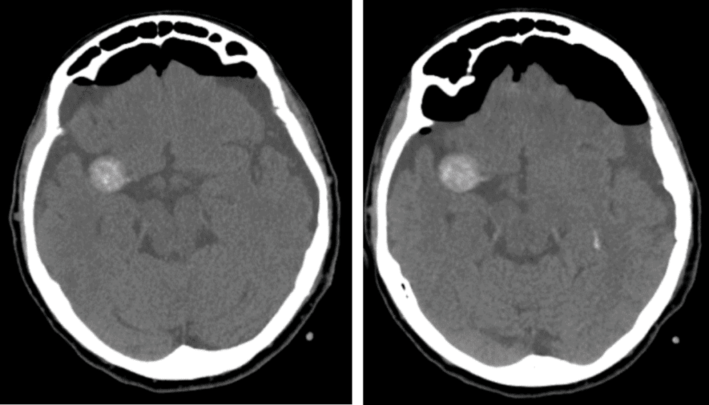
Progressive increase in intracranial pressure over time is life threatening.

Tension Pneumocephalus 

a ct scan of a tension pneumocephalus

Note the progressive compression of the frontal lobes by air with widening of interhemispheric spaces. 

Shared to improve patient safety and healthcare delivery in the provision of radiology services. The circumstances and facts are changed, altered, or deidentified to preserve confidentiality. Privileges have not been waived.

Practical Insights duplicate icon

    • Occurs most commonly after SDH evacuation, but can also be seen after skill base or sinonasal surgeries, head trauma, and nitrous oxide anesthesia.

    • Treated with ventriculostomy, craniotomy, and dural defect closure.


Bell iconTakeaway: Symptomatic enlarging pneumocephalus is a neurosurgical emergency.

The Neuroradiology National Subspecialty Division (NSD) is part of RP’s Clinical Value Team, whichworks to elevate patient care and enhance value through innovation, collaboration and education. To advance this goal, our radiologists and advanced practice providers are committed to sharing peer learning as valuable reminders and insights about what we encounter in our day-to-day practiceCheck back here and on XLinkedIn and Instagram to see these common cases and our findings.

Visit the Clinical Resources page for more cases and to see what we’ve developed to enhance best practice recommendations, elevate image quality and patient care and update current standards throughout RP’s network of practices, all to deliver excellent radiology services to patients, referring clinicians and client partners.

Radiology Partners, through its owned and affiliated practices, is a leading physician-led and physician-owned technology-enabled radiology practice in the U.S. For the latest news from RP, follow us on XLinkedInInstagramYouTube and the blog.


Rad to Rad Learning: Ectopic Pregnancy

The Radiology Partners (RP) Body Imaging Subspecialty Division (NSD) presents our newest Rad to Rad Learning case. 


Peer Learning Opportunitymegaphone icon

This condition affects 1-2% of pregnancies. With associated bleeding, 1st trimester pregnancies are at a risk of up to 18%. 

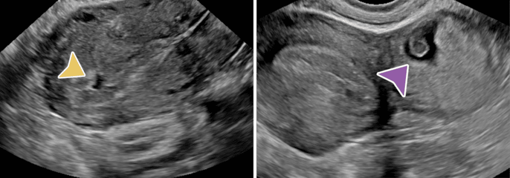
Ectopic Pregnancy93-97% of ectopics are tubal. Tubal ring sign has 95% PPV for ectopic. Visible double decidual sign of the intrauterine sac indicates low probability of ectopic. 

arrows pointing to different types of ectopic pregnancies

Fluid in the endo canal is pseudosac (yellow arrow) vs. Ectopic (purple arrow)

Shared to improve patient safety and healthcare delivery in the provision of radiology services. The circumstances and facts are changed, altered, or deidentified to preserve confidentiality. Privileges have not been waived.


Practical Insights duplicate icon

    • Check for adnexal masses and amount of free fluid in the hepatorenal recess to determine significance.

    • Complex free fluid may represent blood products.

    • Quantitative beta HCG levels are required. Levels >2500 are highly suspicious for occult ectopic.

    • Surgery is recommended for ectopic >3.5cm or if cardiac activity is present.


Bell iconTakeaway: Always report size of ectopic. If >3.5cm, surgery is recommended. 

The Body Imaging Subspecialty Division (NSD) is part of RP’s Clinical Value Team, whichworks to elevate patient care and enhance value through innovation, collaboration and education. To advance this goal, our radiologists and advanced practice providers are committed to sharing peer learning as valuable reminders and insights about what we encounter in our day-to-day practiceCheck back here and on XLinkedIn and Instagram to see these common cases and our findings.

Visit the Clinical Resources page for more cases and to see what we’ve developed to enhance best practice recommendations, elevate image quality and patient care and update current standards throughout RP’s network of practices, all to deliver excellent radiology services to patients, referring clinicians and client partners.

Radiology Partners, through its owned and affiliated practices, is a leading physician-led and physician-owned technology-enabled radiology practice in the U.S. For the latest news from RP, follow us on XLinkedInInstagramYouTube and the blog.


2026 Doctors’ Day: Thank You

March 30 is National Doctors’ Day, and I want to acknowledge the dedication and compassion physicians bring to their work every day in service of patients across the country. 

I am particularly grateful for the radiologists I have the privilege to work alongside and learn from. Together, our radiologists form the foundation of our practice and our mission to transform radiology.  

While much of your impact happens behind the scenes, the reach of your service extends far beyond the reading room. 

You serve our communities in countless ways — from participating in international mission trips to places like Ghana and Guyana, to advocating for thoughtful health policy that strengthens patient care nationwide. You invest in your own growth by engaging in leadership programs, becoming stronger physician leaders for your teams, your practices and our specialty. You lead and mentor the next generation of radiologists, shaping the future of our field, and you advance education and clinical excellence through the important work of our Clinical Value Team. 

Your clinical expertise, your collaborative partnerships with referring providers and care teams, and your unwavering commitment to high-quality radiology care inspire me every day. It’s your work, in all its forms, that brings our mission to life. 

While we continue to navigate challenges across our specialty and the broader healthcare landscape, we remain committed to advancing radiology in innovative, meaningful ways — always with our patients at the center of what we do. 

Thank you for everything you do, for the many ways you serve, and for the difference you make every day. Happy Doctors’ Day! 

Radiology Partners, through its owned and affiliated practices, is a leading physician-led and physician-owned radiology practice in the U.S. Learn more about our mission, values and practice principles at RadPartners.com. For the latest news from RP, follow along on our blog and on X, LinkedIn, Instagram and YouTube. Interested in learning about career opportunities? Visit our careers page


Rad to Rad Learning: Anterior Pneumothorax

The Radiology Partners (RP) Pediatric Radiology National Subspecialty Division (NSD) presents our newest Rad to Rad Learning case.


Peer Learning Opportunitymegaphone icon

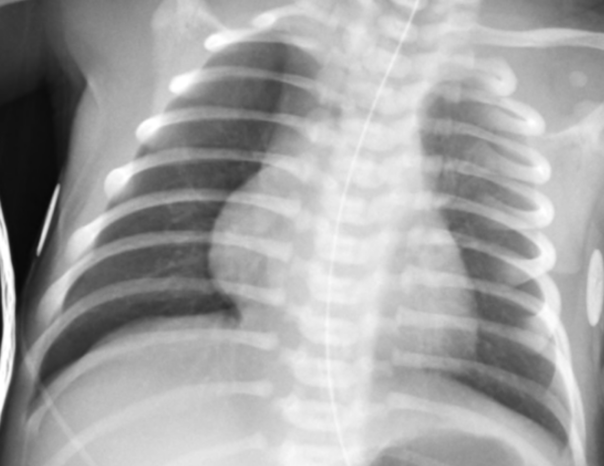
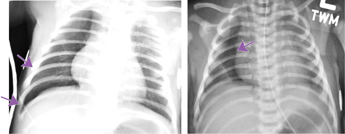
This condition is associated with high morbidity but is difficult to see on supine neonates.

Anterior Pneumothorax

Pediatric chest radiograph

Hallmarks: Deep sulcus sign, no lung marking at the edge of right lung, increased sharpness of the cardiomediastinal border, more prominent on expiration.

Pediatric chest radiograph showing anterior pneumothorax with arrow pointing to landmarksShared to improve patient safety and healthcare delivery in the provision of radiology services. The circumstances and facts are changed, altered, or deidentified to preserve confidentiality. Privileges have not been waived.


Practical Insights duplicate icon

    • May be bilateral.

    • Can occur in both term and preterm babies.

    • Compare lung lucency between both sides.

    • Decubitus radiograph can be a helpful tool for confirmation.


Bell iconTakeaway: Even a suspected pneumothorax is a critical result and should be called.

The Pediatric Radiology National Subspecialty Division (NSD) is part of RP’s Clinical Value Team, whichworks to elevate patient care and enhance value through innovation, collaboration and education. To advance this goal, our radiologists and advanced practice providers are committed to sharing peer learning as valuable reminders and insights about what we encounter in our day-to-day practiceCheck back here and on XLinkedIn and Instagram to see these common cases and our findings.

Visit the Clinical Resources page for more cases and to see what we’ve developed to enhance best practice recommendations, elevate image quality and patient care and update current standards throughout RP’s network of practices, all to deliver excellent radiology services to patients, referring clinicians and client partners.

Radiology Partners, through its owned and affiliated practices, is a leading physician-led and physician-owned technology-enabled radiology practice in the U.S. For the latest news from RP, follow us on XLinkedInInstagramYouTube and the blog.


Rad to Rad Learning: Morton’s Neuroma

The Radiology Partners (RP) MSK National Subspecialty Division (NSD) presents our newest Rad to Rad Learning case.


Peer Learning Opportunitymegaphone icon

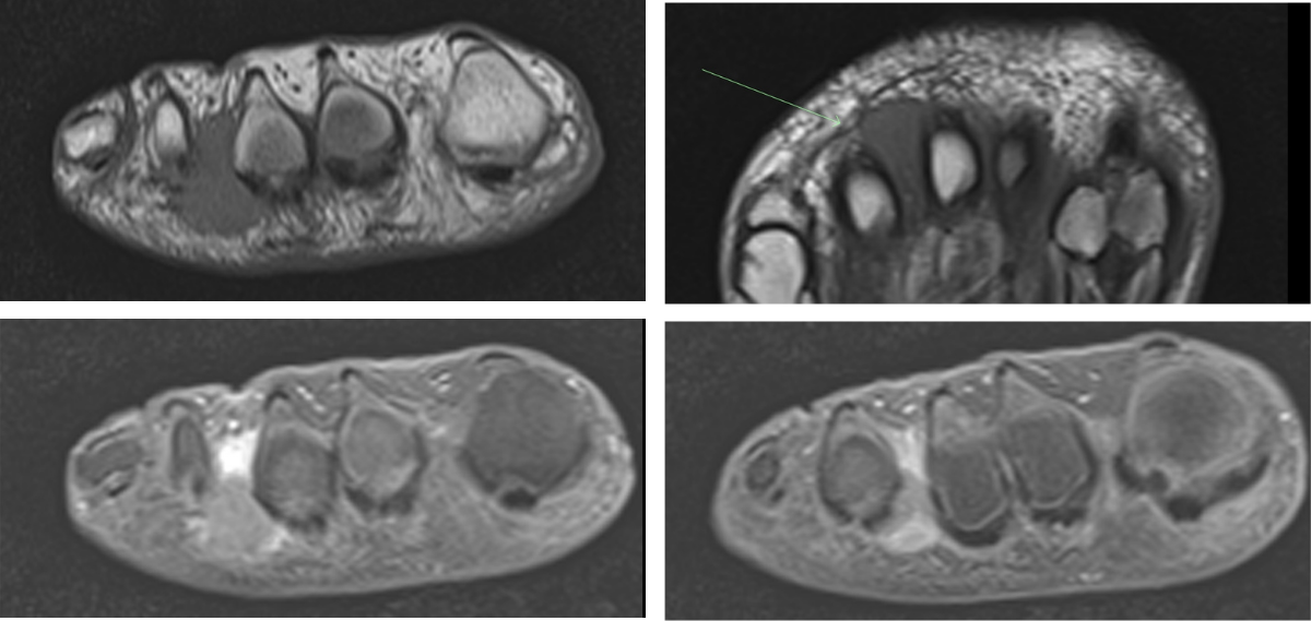
This finding represents a compressive neuropathy of the interdigital nerve primarily in the 2nd and 3rd intermetatarsal spaces.

Morton’s Neuroma

Moron's Neuroma radiographic imageShared to improve patient safety and healthcare delivery in the provision of radiology services. The circumstances and facts are changed, altered, or deidentified to preserve confidentiality. Privileges have not been waived.


Practical Insights duplicate icon

    • Presents with radiating pain, throbbing, numbness, and burning from the webspace into the toes. (Walking on marbles.)

    • Differential diagnosis: fracture, osteonecrosis, bursitis, synovial cyst.

    • Can have concomitant intermetatarsal bursitis.

    • AKA Intermetatarsal perineural fibrosis.


Bell iconTakeaway: Common cause of forefoot pain – best seen on T1 weighted sequences.

The MSK National Subspecialty Division (NSD) is part of RP’s Clinical Value Team, whichworks to elevate patient care and enhance value through innovation, collaboration and education. To advance this goal, our radiologists and advanced practice providers are committed to sharing peer learning as valuable reminders and insights about what we encounter in our day-to-day practiceCheck back here and on XLinkedIn and Instagram to see these common cases and our findings.

Visit the Clinical Resources page for more cases and to see what we’ve developed to enhance best practice recommendations, elevate image quality and patient care and update current standards throughout RP’s network of practices, all to deliver excellent radiology services to patients, referring clinicians and client partners.

Radiology Partners, through its owned and affiliated practices, is a leading physician-led and physician-owned technology-enabled radiology practice in the U.S. For the latest news from RP, follow us on XLinkedInInstagramYouTube and the blog.


Rad to Rad Learning: Slipped Capital Femoral Epiphysis

The Radiology Partners (RP) Pediatric Radiology National Subspecialty Division (NSD) presents our newest Rad to Rad Learning case.


Peer Learning Opportunitymegaphone icon

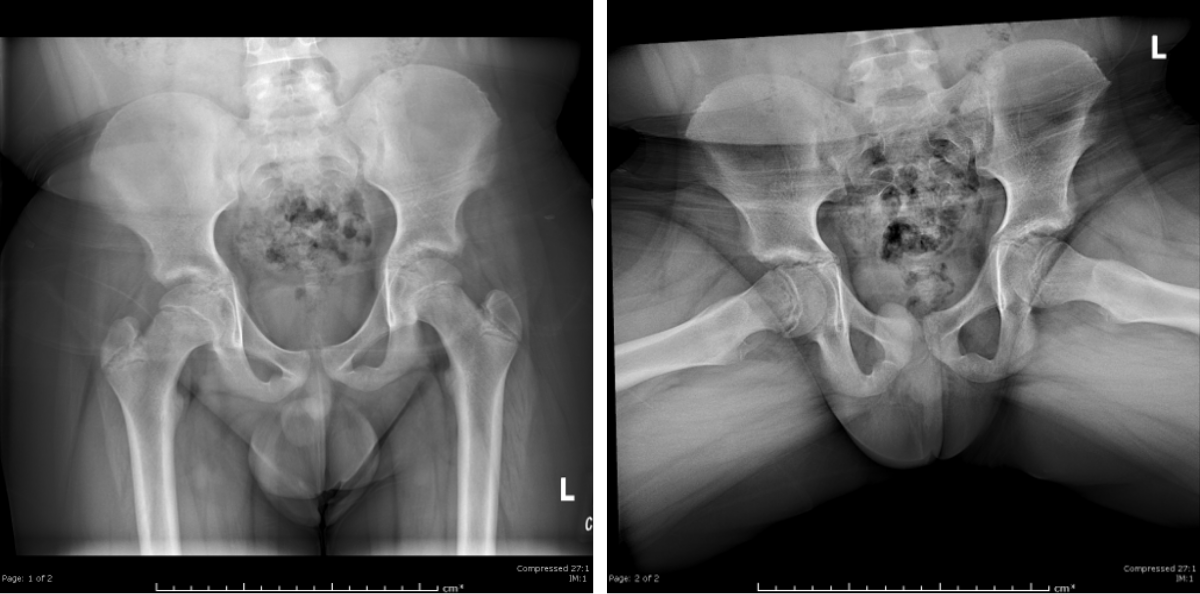
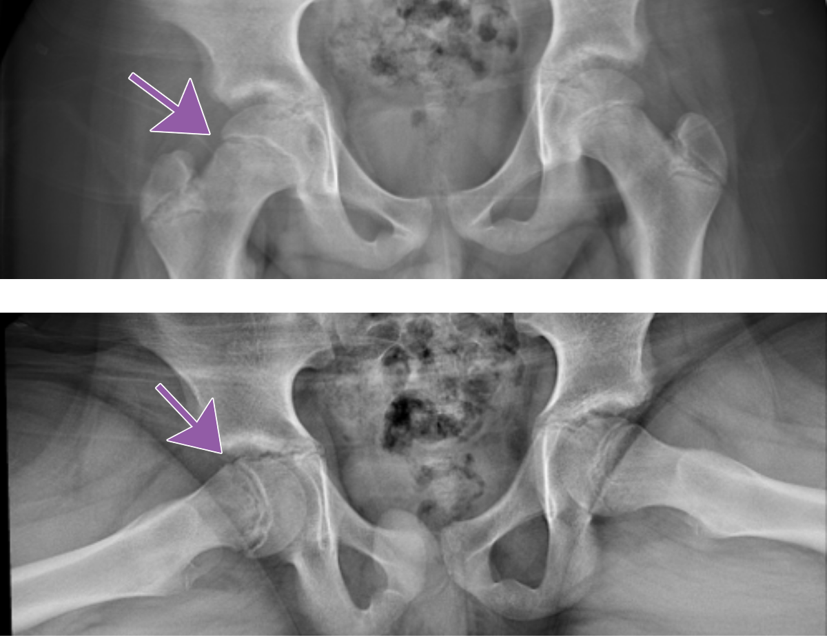
Slipped Capital Femoral Epiphysis (SCFE) is a common and urgent diagnosis and needs to be communicated with the referring provider.

Slipped Capital Femoral Epiphysis

radiographic image of the pelvis

Note the medial and posterior displacement of the right femoral head and widened physis. Frog lateral views are generally more sensitive.

radiographic image of the pelvisShared to improve patient safety and healthcare delivery in the provision of radiology services. The circumstances and facts are changed, altered, or deidentified to preserve confidentiality. Privileges have not been waived.


Practical Insights duplicate icon

    • Commonly missed diagnosis that leads to increased complications.

    • Complications include osteonecrosis, chondrolysis, chronic pain, and growth arrest.

    • Having a high index of suspicion is helpful.

    • For at-risk patients, compare the hips on both views.


Bell iconTakeaway: Make sure to communicate positive findings to the referring provider.

The Pediatric Radiology National Subspecialty Division (NSD) is part of RP’s Clinical Value Team, whichworks to elevate patient care and enhance value through innovation, collaboration and education. To advance this goal, our radiologists and advanced practice providers are committed to sharing peer learning as valuable reminders and insights about what we encounter in our day-to-day practiceCheck back here and on XLinkedIn and Instagram to see these common cases and our findings.

Visit the Clinical Resources page for more cases and to see what we’ve developed to enhance best practice recommendations, elevate image quality and patient care and update current standards throughout RP’s network of practices, all to deliver excellent radiology services to patients, referring clinicians and client partners.

Radiology Partners, through its owned and affiliated practices, is a leading physician-led and physician-owned technology-enabled radiology practice in the U.S. For the latest news from RP, follow us on XLinkedInInstagramYouTube and the blog.


collage of different teammates together

Teammate Appreciation Day 2026: Building the Future of Radiology – Together

At Radiology Partners (RP), our people are the driving force behind everything we do. Every day, our teammates bring purpose, collaboration and a deep commitment to doing what’s right for patients – and that shared spirit continues to propel our mission to transform radiology.

As we recognize National Employee Appreciation Day (or Teammate Appreciation Day, as we call it at RP), this moment offers more than an opportunity to say thank you. It’s also a chance to reflect on the meaningful momentum we’re building together in service of patient care and clinical excellence.

That momentum is evident in the exciting growth across RP over the past year. With the launch of Mosaic Clinical Technologies™ and MosaicOS™, RP is expanding its AI capabilities and clinical services and delivering advanced tools and insights that help care teams work more efficiently and effectively. At the same time, the acquisition of Cognita Imaging Inc. by Mosaic Clinical Technologies™ strengthens our ability to innovate, scale and accelerate solutions that support radiologists, practices and, most importantly, patients.

This collective commitment is also reflected in another achievement worth celebrating: Radiology Partners has earned Great Place to Work® Certification for the sixth consecutive year. More than 80% of RP teammates report that the practice fosters a flexible, welcoming environment where people are trusted with meaningful responsibility and empowered to do their best work.

While individual contributions matter, it’s the collective ingenuity and teamwork across RP that make progress like this possible. From advancements in AI and technology to the daily expertise and compassion of radiologists, technologists and support teams nationwide, everything we do is rooted in service to patients and the communities who rely on us.

Together, RP is building the future of radiology – guided by purpose, powered by people and focused on delivering high-quality, patient-centered care every day.

Thank you to every member of the RP team for all that you do. It is truly an honor to work alongside such a talented, passionate and purpose-driven team!

Radiology Partners, through its owned and affiliated practices, is a leading physician-led and physician-owned radiology practice in the U.S. Learn more about our mission, values and practice principles at RadPartners.com. For the latest news from RP, follow along on our blog and on X, LinkedIn, Instagram and YouTube. Interested in learning about career opportunities? Visit our careers page.