Rad to Rad Learning: Increased Renal Uptake on Bone Scan
The Radiology Partners (RP) NMMI Radiology National Subspecialty Division (NSD)presents our newest Rad to Rad Learning case.
Peer Learning Opportunity
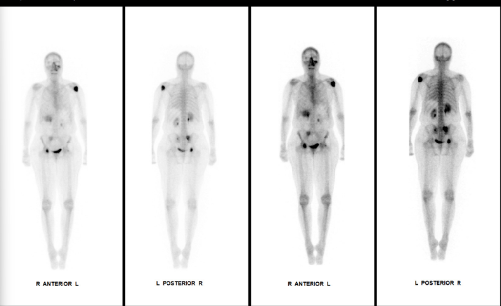
Bone scintigraphy is used in staging and restaging metastatic disease. However, it also provides information regarding renal function in oncology patients.
In the example below, osteoblastic osseous metastases are seen to the pelvic bones and left humerus. Additionally, asymmetric activity is noted to the right kidney in cortical morphology relative to the left kidney.
Shared to improve patient safety and healthcare delivery in the provision of radiology services. The circumstances and facts are changed, altered, or deidentified to preserve confidentiality. Privileges have not been waived.
Practical Insights
Asymmetric unilateral or bilateral renal uptake, also known as “Hot Kidney signs”, typically suggests underlying metabolic or systemic etiologies.
The most common etiology in oncology patients is nephrotoxicity secondary to chemotherapy or other medication.
Other etiologies for this finding include hypercalcemia and cirrhosis.
In rare instances, increased renal uptake can be seen with aluminum breakthrough from radiotracer generator production.
Takeaway: Bone scan provides similar physiologic information on renal function as a renogram.
The NMMI Radiology National Subspecialty Division (NSD) is part of RP’s Clinical Value Team, whichworks to elevate patient care and enhance value through innovation, collaboration and education.To advance this goal, our radiologists and advanced practice providers are committed to sharing peer learning as valuable reminders and insights about what weencounter in our day-to-day practice. Check back here and on X, LinkedIn and Instagram to see these common cases and our findings.
Visit the Clinical Resources page for more cases and to see what we’ve developed to enhance best practice recommendations, elevate image quality and patient care and update current standards throughout RP’s network of practices, all to deliver excellent radiology services to patients, referring clinicians and client partners.
Radiology Partners, through its owned and affiliated practices, is a leading physician-led and physician-owned technology-enabled radiology practice in the U.S. For the latest news from RP, follow us on X, LinkedIn, Instagram, YouTube and the blog.
Rad to Rad Learning: Fat Embolism in Sickle Cell Crisis
The Radiology Partners (RP) Neuroradiology National Subspecialty Division (NSD)presents our newest Rad to Rad Learning case.
Peer Learning Opportunity
Without typical MRI findings Cerebral Fat Embolism can be clinically misdiagnosed as a conventional large vessel stroke or seizure in SCD.
Numerous confluent white matter microinfarcts (Starfield-pattern) and petechial microhemorrhages.
Shared to improve patient safety and healthcare delivery in the provision of radiology services. The circumstances and facts are changed, altered, or deidentified to preserve confidentiality. Privileges have not been waived.
Practical Insights
Occurs in <1% of Sickle Cell Disease (SCD)
Fat globules released due to bone infarcts secondary to vaso-occlusion rather than fracture.
History, distribution of lesions and white matter involvement differentiates from DAI and septic or cardiogenic emboli.
90% of patients recover, but may develop atrophy.
Takeaway: History and MR appearance are crucial for diagnosis.
The Neuroradiology National Subspecialty Division (NSD)is part of RP’s Clinical Value Team, whichworks to elevate patient care and enhance value through innovation, collaboration and education.To advance this goal, our radiologists and advanced practice providers are committed to sharing peer learning as valuable reminders and insights about what weencounter in our day-to-day practice. Check back here and on X, LinkedIn and Instagram to see these common cases and our findings.
Visit the Clinical Resources page for more cases and to see what we’ve developed to enhance best practice recommendations, elevate image quality and patient care and update current standards throughout RP’s network of practices, all to deliver excellent radiology services to patients, referring clinicians and client partners.
Radiology Partners, through its owned and affiliated practices, is a leading physician-led and physician-owned technology-enabled radiology practice in the U.S. For the latest news from RP, follow us on X, LinkedIn, Instagram, YouTube and the blog.
Rad to Rad Learning: High-Risk Aortic Dissection
The Radiology Partners (RP) Interventional Radiology National Subspecialty Division (NSD)presents our newest Rad to Rad Learning case.
Peer Learning Opportunity
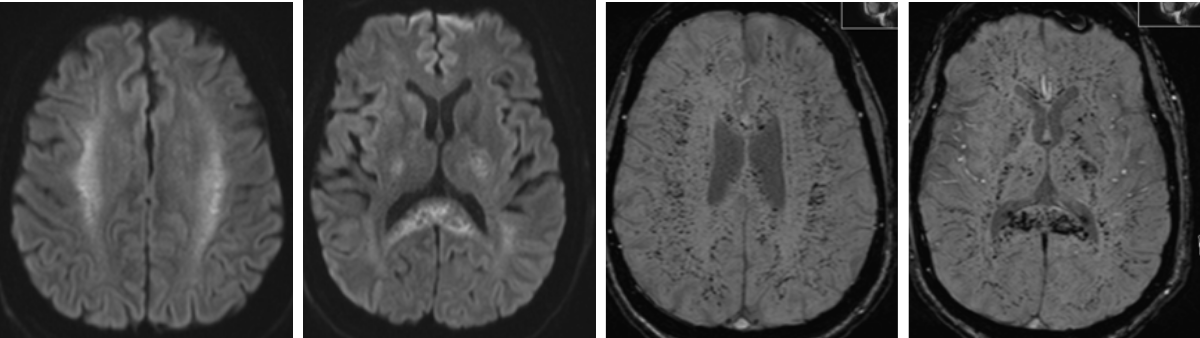
High-risk features should be identified and described to direct management (surgical, endovascular, or hybrid).
Report true / false lumen extension into coronaries, arch vessels, and visceral arteries.
Shared to improve patient safety and healthcare delivery in the provision of radiology services. The circumstances and facts are changed, altered, or deidentified to preserve confidentiality. Privileges have not been waived.
Takeaway: Accurate characterization of dissections improves survival.
The Interventional Radiology National Subspecialty Division (NSD) is part of RP’s Clinical Value Team, whichworks to elevate patient care and enhance value through innovation, collaboration and education.To advance this goal, our radiologists and advanced practice providers are committed to sharing peer learning as valuable reminders and insights about what weencounter in our day-to-day practice. Check back here and on X, LinkedIn and Instagram to see these common cases and our findings.
Visit the Clinical Resources page for more cases and to see what we’ve developed to enhance best practice recommendations, elevate image quality and patient care and update current standards throughout RP’s network of practices, all to deliver excellent radiology services to patients, referring clinicians and client partners.
Radiology Partners, through its owned and affiliated practices, is a leading physician-led and physician-owned technology-enabled radiology practice in the U.S. For the latest news from RP, follow us on X, LinkedIn, Instagram, YouTube and the blog.
Mosaic Clinical Technologies™ Launches Mosaic Reporting™, an AI-Native Reporting Platform Built for Radiology Workflows
Part of MosaicOS, Mosaic Reporting brings a new class of ambient AI, natural dictation and real-time report construction to radiology workflows, helping healthcare organizations expand capacity, reduce reporting friction and move beyond legacy systems.
NASHVILLE, Tenn., June 9, 2026 — Mosaic Clinical Technologies™, a wholly owned subsidiary of Radiology Partners, today announced the commercial launch of Mosaic Reporting™, a new class of AI-native radiology reporting solution available as part of MosaicOS™. Mosaic Reporting is already deployed to thousands of radiologists through Radiology Partners-affiliated practices, giving Mosaic a scaled real-world environment to refine and expand the platform.
Powered by foundation models developed by Cognita Imaging, the AI division of Mosaic Clinical Technologies, Mosaic Reporting is a patent-pending system that helps construct radiology reports in real time as radiologists interpret medical images. Unlike traditional dictation and reporting tools that document findings after a study has been reviewed, Mosaic Reporting works alongside the radiologist during interpretation in real time — organizing findings, structuring reports and enabling targeted edits within the reporting workflow. This capability is made possible by in-house developed foundation models that offer improved speed and accuracy compared to third-party models.
“Radiology is facing a chronic shortage of radiologists as imaging volumes continue to climb, leaving radiology practices, departments and the health systems that depend on them with growing backlogs and turnaround time pressure,” said Mike Peresie, President of Mosaic Clinical Technologies. “Mosaic Reporting is designed to help healthcare organizations create reading capacity, improve turnaround times and better meet service levels that referring physicians and patients expect. Because it is built as part of MosaicOS, organizations can see those gains immediately on a platform that continues to expand.”
Mosaic Reporting™ is purpose-built for radiology workflows and enables:
Real-time, AI-assisted report creation as radiologists dictate
Intelligent extraction of findings from natural speech and automatic placement into appropriate report sections
A natural dictation experience designed to reduce interruptions and cognitive load
Targeted, efficient editing and structured reporting with voice-directed editing
Fewer clicks and automated tasks, allowing radiologists to stay focused on clinical decision-making
Replacement of legacy reporting solutions
“Most AI documentation tools are built around clinician-patient conversations, but radiologists interpret images, which demands a fundamentally different approach,” said Adrit Rao, Technical Product Lead at Cognita Imaging™, the AI division of Mosaic Clinical Technologies. “Mosaic Reporting is one of the first new and significantly advanced approaches to radiology reporting in more than a decade. We worked closely with radiologists to design a reporting experience where AI works in real time as they read, with targeted edits and structured output that reduce friction and keep clinical decision-making at the center.”
For healthcare organizations facing rising imaging demand, staffing constraints, backlogs and pressure to improve turnaround times, Mosaic Reporting is designed to help expand clinical capacity and create a more efficient reporting environment without adding unnecessary complexity.
“This technology creates a more intuitive and effective reporting experience, helping radiologists reduce cognitive burden and focus on the patient,” said Dr. Nina Kottler, Chief Medical AI Officer at Mosaic Clinical Technologies. “For health systems, it creates an opportunity to unlock capacity, improve consistency and build a future-ready foundation that can evolve with their needs.”
About Mosaic Clinical Technologies™ and MosaicOS™
Mosaic Clinical Technologies™, the technology services division of Radiology Partners (RP), is powering the future of radiology through MosaicOS™—a proprietary imaging intelligence platform designed to meet the specialty’s most pressing challenges. A fully cloud-native and AI-native operating system, MosaicOS™ is where innovation meets impact, seamlessly integrating diagnostic technologies, AI-powered tools and smart workflows into a single scalable solution. Mosaic Clinical Technologies™ supports RP’s national network of affiliated practices and commercial partners across the imaging landscape, redefining what is possible in enterprise imaging. Connect with us on LinkedIn and YouTube. Contact us at info@MosaicClinical.ai.
About Radiology Partners
Radiology Partners, through its affiliated practices, is the leading technology-enabled radiology practice in the U.S., serving more than 3,400 hospitals and other healthcare facilities with high quality radiology, technology and artificial intelligence solutions. As a physician-led and physician-owned practice, our mission is to transform radiology by innovating across clinical value, technology, service and economics, while elevating the role of radiology and radiologists in healthcare. Using a proven healthcare services model, Radiology Partners provides consistent, high-quality care to patients, while delivering enhanced value to the hospitals, clinics, imaging centers and referring physicians we serve. Learn more at radpartners.com and connect with us on LinkedIn, X, Instagram and YouTube. Talented radiologists who want to practice with industry leading, cutting-edge tools should reach out to us at recruiting@radpartners.com.
Marie Currie, a multimedia artist, never expected a persistent cough to change the course of her life. As she focused on her craft, the cough wouldn’t go away, so she sought answers.
After extensive testing, she was diagnosed with pulmonary fibrosis, a serious condition that came with an uncertain future. Although the diagnosis was difficult, it set in motion a series of events that would ultimately save her life. As part of her care, Marie underwent routine imaging every few months to monitor the progression of her condition. During one of these scans, physicians identified a small abnormality — a tiny spot that would later prove critical.
When Marie’s care team noticed the lesion growing, she was referred to Dr. Sameer Rehman, a cardiothoracic and interventional radiologist at Desert Radiology in Las Vegas, Nevada, which is affiliated with Radiology Partners (RP). He specializes in advanced imaging of the heart and lungs and minimally invasive image-guided procedures, with focused special interest in diagnosing and treating lung cancer with thermal ablation. Marie’s underlying lung disease would make surgery risky, and after evaluating her case, Dr. Rehman recommended a minimally invasive approach: a combined biopsy and lung cryoablation procedure.
During the procedure, a biopsy confirmed the presence of lung cancer. In the same session, Dr. Rehman performed a cryoablation procedure, which involves the use of CT guidance to precisely place cryoablation probes into the tumor, creating an “ice ball” around the tumor and effectively destroying it while preserving surrounding lung function.
This approach reflects a growing shift in cancer care: using targeted, minimally invasive therapies to treat disease earlier, with fewer complications and faster recovery. For patients with underlying conditions like pulmonary fibrosis and emphysema, these options can be especially impactful.
Marie’s outcome was remarkable. Follow-up imaging at six months and one year showed no evidence of active cancer, only a small residual scar where the tumor had been treated.
“She has been cancer-free post-ablation now and has been doing clinically well,” Dr. Rehman said.
For Marie, the impact goes far beyond the medical result.
“I cannot stress enough to people that are in my situation that they need to know that this option is available for them, and I couldn’t be doing better,” she said. “I owe that procedure and his expertise my life, and I don’t believe that I would be here long for my grandson if it wasn’t for how blessed I was that this procedure was available at the time it was. And boy do I love being a grandma.”
Radiology Partners, through its owned and affiliated practices, is a leading physician-led and physician-owned radiology practice in the U.S. Learn more about our mission, values and practice principles at RadPartners.com. For the latest news from RP, follow along on our blog and on X, LinkedIn, Instagram and YouTube. Interested in learning about career opportunities? Visit our careers page.
PLS 2026: Congratulations to Our Award Winners
At our annual Practice Leadership Summit (PLS), we take time to celebrate Radiology Partners (RP) teammates who live our values in action.
Year after year, this tradition gives us the opportunity to recognize those who demonstrate integrity, teamwork, excellence, service and accountability in how they show up for others. Their example inspires all of us to grow, both as colleagues and as individuals. By honoring these teammates, we keep our values front and center as we continue advancing our mission to transform radiology.
The three 2026 Value Award winners were nominated by their peers and selected through a rigorous process, and we are thankful they are on our team. Congratulations!
Service: We exist to provide the best in radiology services. We strive to understand the needs of our clients – especially patients and referring physicians – and exceed their expectations.
Kati Brady: Service Award
Kati Brady serves as Vice President, Managing General Counsel at RP.
Colleagues describe Kati as someone who consistently delivers service through her steady presence, focus and intentional support of others. She is known for navigating complex, high‑stakes situations with clarity and care, ensuring people feel heard, supported and confident in the path forward. Kati brings insight beyond her role, offers thoughtful solutions when challenges feel stuck, and does so with warmth and humility. By showing up with consistency and authenticity, she exemplifies service not just through what she does but how she makes others feel, and in doing so, elevates those around her.
Teamwork: The core of our Practice. We work together. The best of us alone cannot exceed the impact of all of us together. We support, respect and value each other.
Dr. Steve Craig: Teamwork Award
Dr. Steve Craig is the Associate Chief Medical Officer (ACMO) of Operations and Integrations and Practice President of the RP SEAL Team.
Dr. Craig was recognized for his exceptional teamwork during times of significant change. Known as a master of diagnostic radiology with deep interventional expertise, he leads with calm confidence and an unwavering focus on patient care. Colleagues describe him as someone who can always be counted on: ready to step in, travel where needed and help solve challenges that may seem impossible. Whether guiding high‑stakes go‑lives or supporting teams behind the scenes, Dr. Craig creates trust, steady leadership and a shared belief in success, exemplifying what teamwork looks like at RP.
Accountability: Driven by our desire for continuous improvement. We take responsibility for our actions and acknowledge that each of us has a role in the success of the practice.
Dr. Vik Krishnasetty: Accountability Award
Dr. Vik Krishnasetty serves as Associate Chief Medical Officer for Clinical Technology and Data.
Colleagues describe Dr. Vik Krishnasetty as a leader who consistently advances radiology through thoughtful innovation, operational rigor, and unwavering commitment to improvement. Throughout his career, he has worked to strengthen clinical workflows, leading teams to make bold changes that remove barriers and better define the value of radiologists’ work. He is known as a true partner across functions, contributing wherever the need is greatest—whether solving complex operational challenges, developing smarter systems or helping teams adopt new capabilities.
Chairman’s Impact Award
Zhihong Chen: Chairman’s Impact Award
RP announced a new award this year, the Chairman’s Impact Award. The award recognizes individuals whose contributions have had an exceptional impact on the practice, often behind the scenes, and whose efforts meaningfully advance the mission and future of RP.
Zhihong Chen, Cognita Co-founder and Chief Technology Officer, was honored for his transformative work using technology to advance the future of RP, radiology and, more broadly, healthcare. Cognita—now part of Mosaic Clinical Technologies, RP’s technology and AI services division—plays a key role in bringing advanced AI capabilities into RP’s clinical workflows, helping scale innovation across the practice.
Zhihong combines deep expertise in AI with a relentless work ethic and a clear, mission-driven focus on improving patient care. Colleagues highlighted his ability to turn complex clinical challenges into practical solutions, inspire those around him, and elevate the work of the entire team—all while leading with humility and a genuine passion for making a difference.
Please join us in congratulating the 2026 Value Award recipients! Learn more about RP’s values on our website or on our YouTube channel.
Radiology Partners, through its owned and affiliated practices, is a leading physician-led and physician-owned technology-enabled radiology practice in the U.S. Learn more about our mission, values and practice principles at RadPartners.com. For the latest news from RP, follow along on our blog and on X, LinkedIn, Instagram and YouTube. Interested in learning about career opportunities? Visit our careers page.
Rad Reach: Strengthening Communities Through Partnership in Ghana
In late 2025, Radiology Partners (RP) returned to Ghana, Africa for an international service trip with a shared commitment to partnership, sustainability and service.
Building on a long-standing collaboration with RAD-AID International and the support provided during RP’s 2024 trip to Ghana, this year’s mission expanded in both scope and impact – bringing together clinical and support teammates to serve hospitals, schools and entrepreneurs across Accra, Kumasi and neighboring communities.
Dr. Arthy Saravanan, RP Associate Chief Medical Officer for Recruitment, explained the purpose was twofold: to continue meaningful work already underway and to create a broader, more lasting impact. “We wanted to create a larger impact this time, so we took a larger team with us, both on the clinical and the support teammate side.”
Strengthening Clinical Care Through Collaboration
A 17-member clinical team of RP radiologists and technologists served four hospitals across two cities, working alongside local physicians, residents and technologists. The focus extended beyond service delivery to education, mentorship and systems that support long-term quality care.
For many participants, returning to Ghana was essential to ensuring sustainability. “We don’t want to just go into a place, give and then leave – leaving them back where they started,” said Dr. Arlene Richardson, musculoskeletal radiologist. “Building on the previous trips definitely shows how this outreach can be sustainable and have long-term impact.”
Throughout the week, RP clinicians shared hands-on skills, developed reporting templates, refined imaging techniques and collaborated on complex cases. The enthusiasm and engagement of local trainees stood out.
“They’re very eager to learn,” said Dr. Leonardo Freitas, neuroradiologist. “It was priceless to me. I’ll never forget this experience.”
RP teammates came together to ensure patients received needed care – sharing equipment, supplies and expertise. Dr. Shambhavi Venkataraman, breast imaging radiologist, recalled the first day in Ghana, when the RP clinical team planned to teach radiology fellows how to do biopsies. “We realized they have no equipment – they have no biopsy needles.” The RP team had brought supplies and was able to provide the needed biopsy equipment. “That just shows the RP spirit,” said Dr. Venkataraman. “We’re all like, ‘Your patients need them. You take them.’”
Investing in Education and Technology
Alongside the clinical mission, a team of four RP support teammates focused on expanding access to technology in local schools. Building on last year’s work, the team established new technology labs at two schools in Aboasa and Gyakiti and revisited a previously served site in Akwamufie to extend the life of existing equipment.
The work required creativity and flexibility, from refurbishing aging computers to adapting installations in real time. But the impact was immediate and visible. “When the kids come into the computer lab, playing these educational games, and smiling and crowding around one another in order to interact with technology on a level they weren’t able to before – that’s where you really feel like we’re doing something there,” said Scott Kozel, Systems Engineer.
Beyond the equipment, the team emphasized relationships, values and shared experience. “We didn’t just go there to complete a mission,” said Rebekah Thompson, Director of IT Portfolio Management, adding that the team embedded RP’s values – integrity, teamwork, excellence, service and accountability – into everything they did. “The relationships that we built, fostering ongoing collaboration and support, and then just the joy that was shared…having the ability to take the time out, to interact, to have fun, experience and engage with the kids was so meaningful. We’re touching so many lives.”
Empowering Entrepreneurs and Communities
A third group of RP leaders led an entrepreneurship master class in Accra, partnering with local business owners and aspiring entrepreneurs. The sessions focused on practical skills – finance, marketing, communication and business sustainability – while honoring the deep connection between entrepreneurship and community impact.
Participants brought diverse backgrounds and strong ambitions, creating a highly collaborative learning environment. “They’re already entrepreneurs. They have thriving businesses,” said Jenny Benoit, Vice President of Practice Finance.
“Everything that they were doing had some connection to service in the community,” said Sarah Mahlstedt, Vice President of Strategic Finance and Operational Systems. “Even something that just felt like basic commerce always was connected back to the community. So being able to learn more and add whatever value we could to their businesses, it really did feel like we were contributing to the broader Accra community and delivering something of value.”
For many RP participants, the experience reinforced the broader purpose. “These types of missions remind us of the importance of service,” said Jâlie Cohen, chief human resources officer.
A Lasting Ripple Effect
For the RP team, the trip to Ghana reflected the power of partnership grounded in humility, expertise and shared purpose. “I’ve never been more proud of being in RP than when I went to Ghana,” said Dr. Frank Castellano, Associate Chief Medical Officer of Clinical Operations. “That collective group of people going with the mentality of just being there to help and teach was really special.”
Radiology Partners, through its affiliated practices, is a leading physician-led and physician-owned technology-enabled radiology practice in the U.S. Learn more about our mission, values and practice principles at RadPartners.com. For the latest news from RP, follow along on our blog and on X, LinkedIn, Instagram and YouTube. Interested in learning about career opportunities? Visit our careers page.
Clinical Pathway: RP’s Clinical Value Team presents best practices for stroke CT perfusion interpretation
In stroke care, imaging decisions can change everything.
The Clinical Pathway gives radiologists a clear framework for applying perfusion imaging in real-world stroke cases, from identifying potentially salvageable brain tissue to recognizing technical limitations, interpretation pitfalls and cases where automated outputs need a second look.
At its core, this is about using advanced imaging wisely: AI can assist and perfusion maps can guide, but radiologist judgment still drives the interpretation. Fast is good. Accurate is better. Both are the goal.
This Clinical Pathway reflects the expertise and practical insight of our Neuroradiology NSD, and we’re grateful for their leadership in advancing stroke imaging education across the practice.
The Neuroradiology NSD and its advisory board is made up of practicing radiologists spearhead the development and implementation of programs with a mission to enhance clinical value and quality in imaging across RP. They focus on refining best practice recommendations, advancing image quality and aligning with the latest industry standards, all to deliver innovation and excellence in radiology services for patients, referring clinicians and client partners, and they share resources, like this clinical pathway, broadly so that all practices can deliver high-quality subspecialty care to patients in their communities.
Radiology Partners Clinical Value Team exists to elevate patient care and enhance value through innovation, collaboration and education. Radiology Partners, through its owned and affiliated practices, is a leading physician-led and physician-owned radiology practice in the U.S. For the latest news from RP, follow us on X, LinkedIn, Instagram, YouTube and the blog.
Shared to improve patient safety and healthcare delivery in the provision of radiology services. The circumstances and facts are changed, altered, or deidentified to preserve confidentiality. Privileges have not been waived.
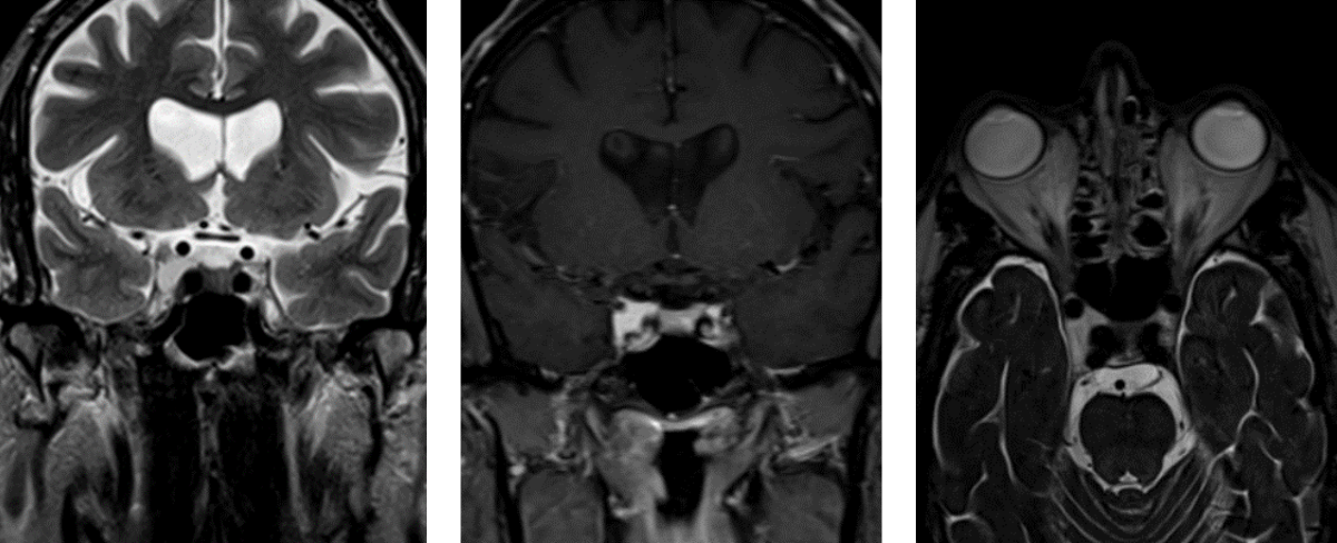
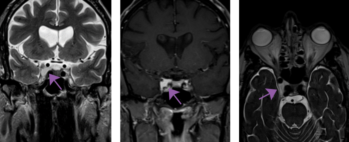
Rad to Rad Learning: Cavernous Sinus Hemangioma
The Radiology Partners (RP) Neuroradiology National Subspecialty Division (NSD) presents our newest Rad to Rad Learning case.
Peer Learning Opportunity
High risk of intraoperative hemorrhage with over 10% mortality rate if surgical removal is attempted.
Shared to improve patient safety and healthcare delivery in the provision of radiology services. The circumstances and facts are changed, altered, or deidentified to preserve confidentiality. Privileges have not been waived.
Practical Insights
Marked T2 hyperintensity, vascular encasement, progressive “filling-in”, vascular blush, or the absence of hyperostosis, vascular narrowing, or connection to the pituitary gland differentiates it from meningioma, adenoma, schwannoma, or metastasis.
Takeaway: : Pre-treatment diagnosis is critical since lesion is radiosensitive, but surgically challenging.
The Neuroradiology National Subspecialty Division (NSD) ) is part of RP’s Clinical Value Team, whichworks to elevate patient care and enhance value through innovation, collaboration and education.To advance this goal, our radiologists and advanced practice providers are committed to sharing peer learning as valuable reminders and insights about what weencounter in our day-to-day practice. Check back here and on X, LinkedIn and Instagram to see these common cases and our findings.
Visit the Clinical Resources page for more cases and to see what we’ve developed to enhance best practice recommendations, elevate image quality and patient care and update current standards throughout RP’s network of practices, all to deliver excellent radiology services to patients, referring clinicians and client partners.
Radiology Partners, through its owned and affiliated practices, is a leading physician-led and physician-owned technology-enabled radiology practice in the U.S. For the latest news from RP, follow us on X, LinkedIn, Instagram, YouTube and the blog.
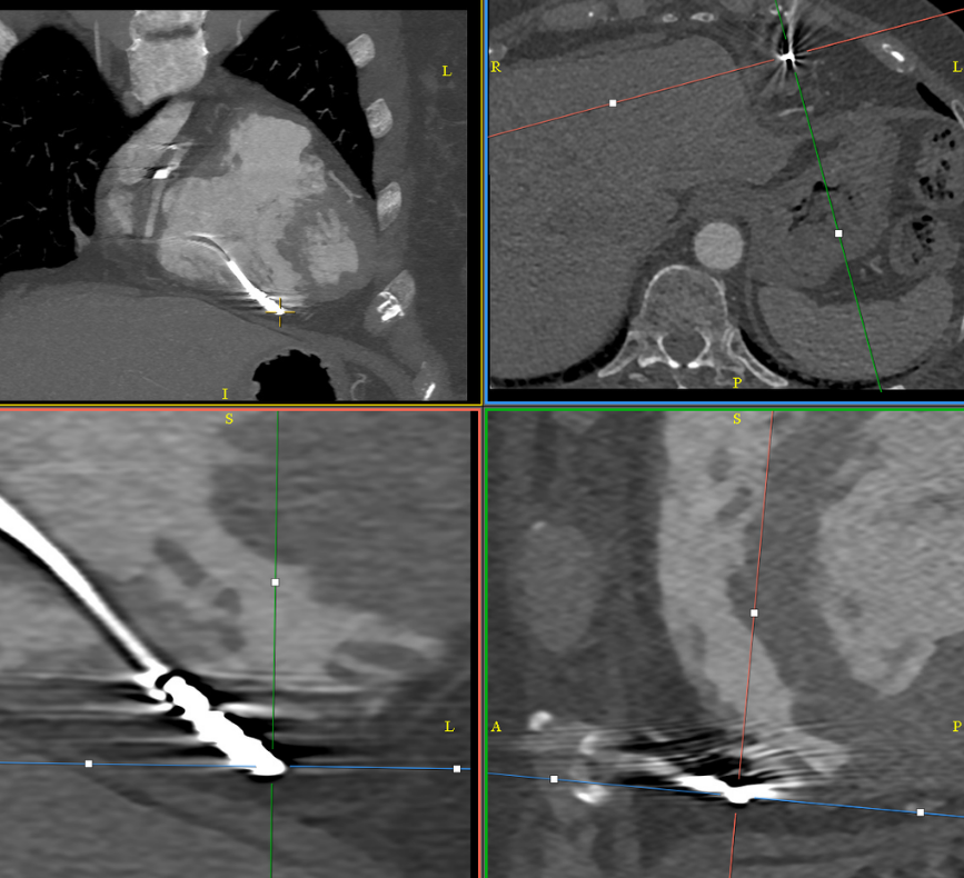
Rad to Rad Learning: Perforated Pacer Lead
The Radiology Partners (RP) Cardiothoracic Imaging National Subspecialty Division (NSD)presents our newest Rad to Rad Learning case.
Peer Learning Opportunity
This patient has a normally functioning pacemaker, but where is the tip of this pacer lead?
Artifacts present in standard axial views can make it difficult to localalize the tips of pacer leads. Oblique MPRs should be used to clarify positioning.
Shared to improve patient safety and healthcare delivery in the provision of radiology services. The circumstances and facts are changed, altered, or deidentified to preserve confidentiality. Privileges have not been waived.
Practical Insights
Even though pacer lead complications are rare, pacemakers are common devices.
Devices may be functional even with perforation, but may result in complications on attempted lead placement.
Takeaway: Lead perforation may not be clinically evident, but can be detected by imaging.
The Cardiothoracic Imaging National Subspecialty Division (NSD) is part of RP’s Clinical Value Team, whichworks to elevate patient care and enhance value through innovation, collaboration and education.To advance this goal, our radiologists and advanced practice providers are committed to sharing peer learning as valuable reminders and insights about what weencounter in our day-to-day practice. Check back here and on X, LinkedIn and Instagram to see these common cases and our findings.
Visit the Clinical Resources page for more cases and to see what we’ve developed to enhance best practice recommendations, elevate image quality and patient care and update current standards throughout RP’s network of practices, all to deliver excellent radiology services to patients, referring clinicians and client partners.
Radiology Partners, through its owned and affiliated practices, is a leading physician-led and physician-owned technology-enabled radiology practice in the U.S. For the latest news from RP, follow us on X, LinkedIn, Instagram, YouTube and the blog.


 Shared to improve patient safety and healthcare delivery in the provision of radiology services. The circumstances and facts are changed, altered, or deidentified to preserve confidentiality. Privileges have not been waived.
 Takeaway: Bone scan provides similar physiologic information on renal function as a renogram.
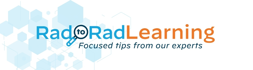
 Shared to improve patient safety and healthcare delivery in the provision of radiology services. The circumstances and facts are changed, altered, or deidentified to preserve confidentiality. Privileges have not been waived.


 Shared to improve patient safety and healthcare delivery in the provision of radiology services. The circumstances and facts are changed, altered, or deidentified to preserve confidentiality. Privileges have not been waived.
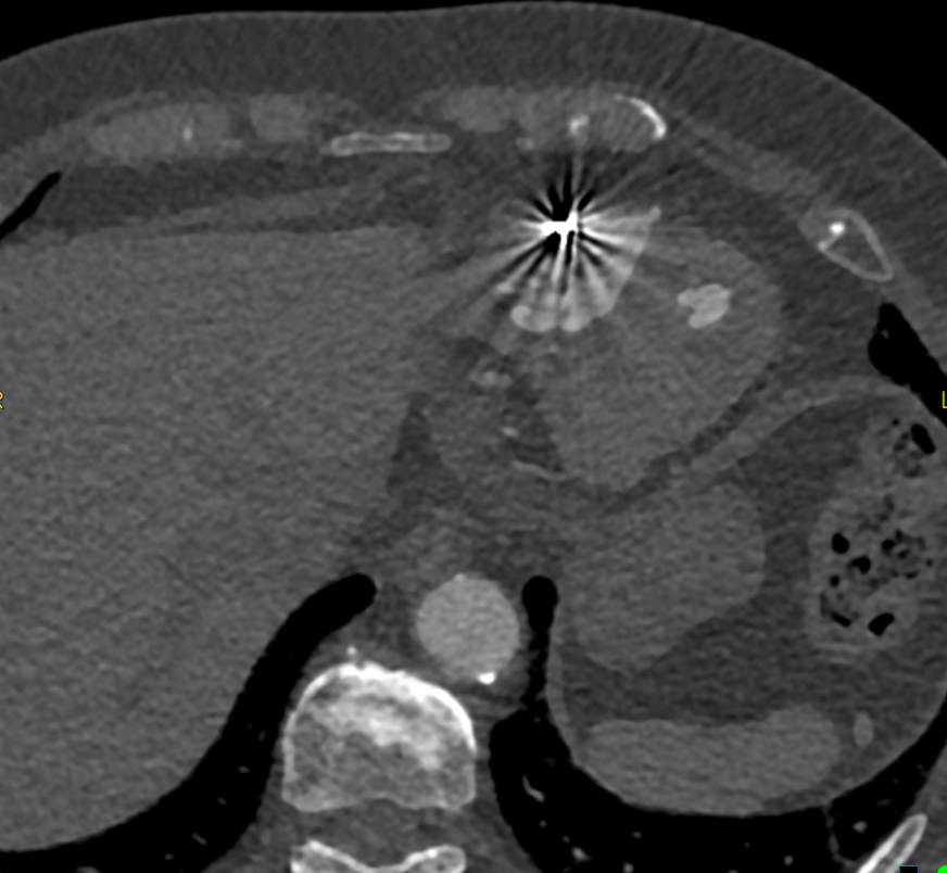
 Shared to improve patient safety and healthcare delivery in the provision of radiology services. The circumstances and facts are changed, altered, or deidentified to preserve confidentiality. Privileges have not been waived.