The Radiology Partners (RP) Body Imaging National Subspecialty Division (NSD) presents our newest Rad to Rad Learning case.
Peer Learning Opportunity
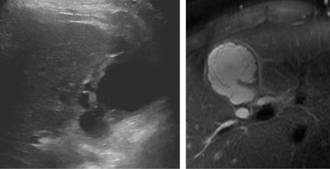
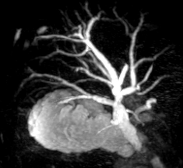
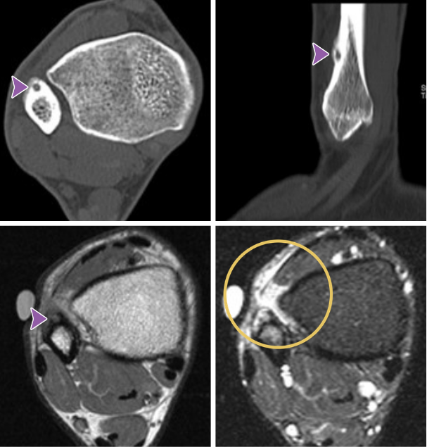
Around 2% of patients with acute cholecystitis will develop gangrenous cholecystitis, which has an elevated risk of perforation and a 22% mortality rate.
Ultrasound features include gall bladder distension measuring greater than 4 cm in short axis, wall disruption, mucosal irregularity, intraluminal membranes and pericholecystic fluid.
 Shared to improve patient safety and healthcare delivery in the provision of radiology services. The circumstances and facts are changed, altered, or deidentified to preserve confidentiality. Privileges have not been waived.
Practical Insights 
-
- Clinical manifestations include findings of sepsis and severe right upper right quadrant pain.
- Murphy’s sign may be absent in 2/3 of patients.
- Risk factors include male sex, diabetes, age >45, and cardiovascular disease.
- Urgent cholecystectomy is the appropriate management.
 Takeaway: The hallmark is mucosal sloughing.
The Body Imaging National Subspecialty Division (NSD) is part of RP’s Clinical Value Team, which works to elevate patient care and enhance value through innovation, collaboration and education. To advance this goal, our radiologists and advanced practice providers are committed to sharing peer learning as valuable reminders and insights about what we encounter in our day-to-day practice. Check back here and on X, LinkedIn and Instagram to see these common cases and our findings.
Visit the Clinical Resources page for more cases and to see what we’ve developed to enhance best practice recommendations, elevate image quality and patient care and update current standards throughout RP’s network of practices, all to deliver excellent radiology services to patients, referring clinicians and client partners.
Radiology Partners, through its owned and affiliated practices, is a leading physician-led and physician-owned technology-enabled radiology practice in the U.S. For the latest news from RP, follow us on X, LinkedIn, Instagram, YouTube and the blog.

 Shared to improve patient safety and healthcare delivery in the provision of radiology services. The circumstances and facts are changed, altered, or deidentified to preserve confidentiality. Privileges have not been waived.
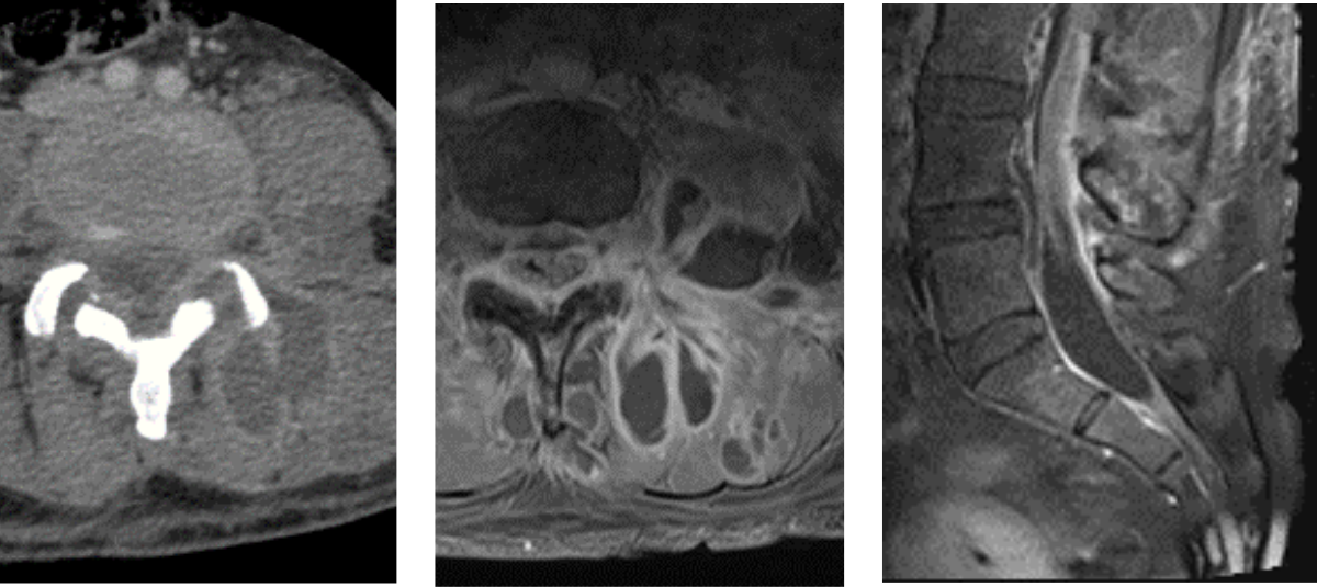 Shared to improve patient safety and healthcare delivery in the provision of radiology services. The circumstances and facts are changed, altered, or deidentified to preserve confidentiality. Privileges have not been waived.
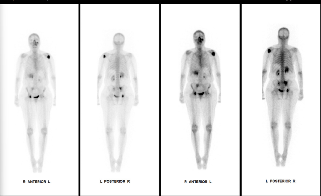 Shared to improve patient safety and healthcare delivery in the provision of radiology services. The circumstances and facts are changed, altered, or deidentified to preserve confidentiality. Privileges have not been waived.
 Shared to improve patient safety and healthcare delivery in the provision of radiology services. The circumstances and facts are changed, altered, or deidentified to preserve confidentiality. Privileges have not been waived.

 Shared to improve patient safety and healthcare delivery in the provision of radiology services. The circumstances and facts are changed, altered, or deidentified to preserve confidentiality. Privileges have not been waived.