A doctor with a stethoscope around his neck stands next to a smiling woman in an RP clinic hallway, both looking at the camera. The image is faded and softly lit.

Patient Save: Marie Currie’s story

Marie Currie, a multimedia artist, never expected a persistent cough to change the course of her life. As she focused on her craft, the cough wouldn’t go away, so she sought answers.

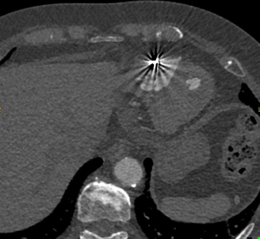
After extensive testing, she was diagnosed with pulmonary fibrosis, a serious condition that came with an uncertain future. Although the diagnosis was difficult, it set in motion a series of events that would ultimately save her life. As part of her care, Marie underwent routine imaging every few months to monitor the progression of her condition. During one of these scans, physicians identified a small abnormality — a tiny spot that would later prove critical.

When Marie’s care team noticed the lesion growing, she was referred to Dr. Sameer Rehman, a cardiothoracic and interventional radiologist at Desert Radiology in Las Vegas, Nevada, which is affiliated with Radiology Partners (RP). He specializes in advanced imaging of the heart and lungs and minimally invasive image-guided procedures, with focused special interest in diagnosing and treating lung cancer with thermal ablation. Marie’s underlying lung disease would make surgery risky, and after evaluating her case, Dr. Rehman recommended a minimally invasive approach: a combined biopsy and lung cryoablation procedure.

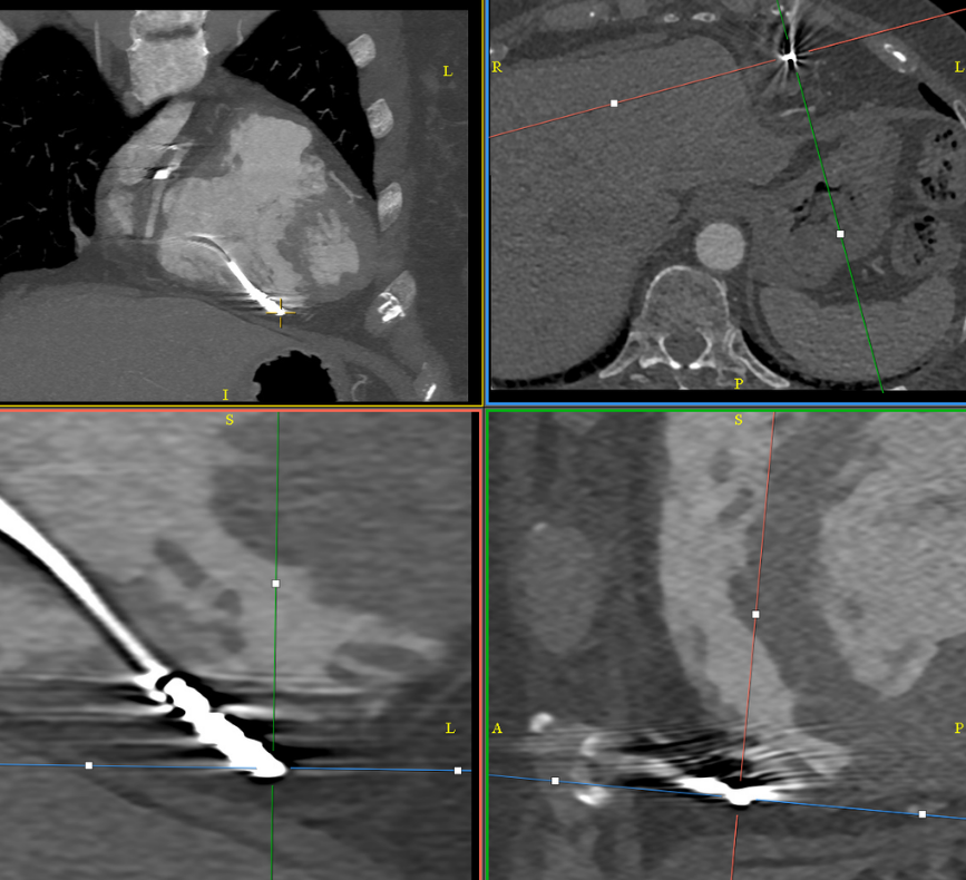
During the procedure, a biopsy confirmed the presence of lung cancer. In the same session, Dr. Rehman performed a cryoablation procedure, which involves the use of CT guidance to precisely place cryoablation probes into the tumor, creating an “ice ball” around the tumor and effectively destroying it while preserving surrounding lung function.

This approach reflects a growing shift in cancer care: using targeted, minimally invasive therapies to treat disease earlier, with fewer complications and faster recovery. For patients with underlying conditions like pulmonary fibrosis and emphysema, these options can be especially impactful.

Marie’s outcome was remarkable. Follow-up imaging at six months and one year showed no evidence of active cancer, only a small residual scar where the tumor had been treated.

“She has been cancer-free post-ablation now and has been doing clinically well,” Dr. Rehman said.

For Marie, the impact goes far beyond the medical result.

“I cannot stress enough to people that are in my situation that they need to know that this option is available for them, and I couldn’t be doing better,” she said. “I owe that procedure and his expertise my life, and I don’t believe that I would be here long for my grandson if it wasn’t for how blessed I was that this procedure was available at the time it was. And boy do I love being a grandma.”

Read more about lung cryoablation in this white paper co-authored by Dr. Rehman, “Lung Ablation Outcomes for Inoperable Stage IA Non-Small Cell Lung Cancer,” which was published in the Journal of Vascular and Interventional Radiology in March 2026.

Radiology Partners, through its owned and affiliated practices, is a leading physician-led and physician-owned radiology practice in the U.S. Learn more about our mission, values and practice principles at RadPartners.com. For the latest news from RP, follow along on our blog and on XLinkedInInstagram and YouTube. Interested in learning about career opportunities? Visit our careers page.


A dark blue background with various hexagonal shapes and lines clustered on the left side, representing rad to rad learning, gradually fading into open space on the right.

Rad to Rad Learning: Perforated Pacer Lead

The Radiology Partners (RP) Cardiothoracic Imaging National Subspecialty Division (NSD) presents our newest Rad to Rad Learning case. 


Peer Learning Opportunitymegaphone icon

This patient has a normally functioning pacemaker, but where is the tip of this pacer lead?

Artifacts present in standard axial views can make it difficult to localalize the tips of pacer leads. Oblique MPRs should be used to clarify positioning.

Perforated Pacer LeadPerforated Pacer Lead with arrows pointing to landmarksShared to improve patient safety and healthcare delivery in the provision of radiology services. The circumstances and facts are changed, altered, or deidentified to preserve confidentiality. Privileges have not been waived.


Practical Insights duplicate icon

    • Even though pacer lead complications are rare, pacemakers are common devices.

    • Devices may be functional even with perforation, but may result in complications on attempted lead placement.


Bell iconTakeaway: Lead perforation may not be clinically evident, but can be detected by imaging.

The Cardiothoracic Imaging National Subspecialty Division (NSD) is part of RP’s Clinical Value Team, whichworks to elevate patient care and enhance value through innovation, collaboration and education. To advance this goal, our radiologists and advanced practice providers are committed to sharing peer learning as valuable reminders and insights about what we encounter in our day-to-day practiceCheck back here and on XLinkedIn and Instagram to see these common cases and our findings.

Visit the Clinical Resources page for more cases and to see what we’ve developed to enhance best practice recommendations, elevate image quality and patient care and update current standards throughout RP’s network of practices, all to deliver excellent radiology services to patients, referring clinicians and client partners.

Radiology Partners, through its owned and affiliated practices, is a leading physician-led and physician-owned technology-enabled radiology practice in the U.S. For the latest news from RP, follow us on XLinkedInInstagramYouTube and the blog.


A dark blue background with various hexagonal shapes and lines clustered on the left side, representing rad to rad learning, gradually fading into open space on the right.

Rad to Rad Learning: Mode of Communication

The Radiology Partners (RP) Cardiothoracic Imaging National Subspecialty Division (NSD) presents our newest Rad to Rad Learning case.


Peer Learning Opportunitymegaphone icon

This is a routine outpatient chest x-ray that just showed up on your list at 4:45 pm on a Friday. What is the appropriate management?

Mode of Communication

Chest X-rayShared to improve patient safety and healthcare delivery in the provision of radiology services. The circumstances and facts are changed, altered, or deidentified to preserve confidentiality. Privileges have not been waived.


Practical Insights duplicate icon

    • Call the referring provider right away. Don’t risk the report sitting in their inbox until Monday.

    • We’re all familiar with critical findings that need to be communicated immediately like strokes or PEs, but it is also important to communicate more “routine” findings in a way that would prevent delays in treatment that could cause significant adverse outcomes.


Bell iconTakeaway: Consider potential treatment delays in how you choose to communicate your findings.

The Cardiovascular Imaging National Subspecialty Division (NSD) is part of RP’s Clinical Value Team, whichworks to elevate patient care and enhance value through innovation, collaboration and education. To advance this goal, our radiologists and advanced practice providers are committed to sharing peer learning as valuable reminders and insights about what we encounter in our day-to-day practiceCheck back here and on XLinkedIn and Instagram to see these common cases and our findings.

Visit the Clinical Resources page for more cases and to see what we’ve developed to enhance best practice recommendations, elevate image quality and patient care and update current standards throughout RP’s network of practices, all to deliver excellent radiology services to patients, referring clinicians and client partners.

Radiology Partners, through its owned and affiliated practices, is a leading physician-led and physician-owned technology-enabled radiology practice in the U.S. For the latest news from RP, follow us on XLinkedInInstagramYouTube and the blog.


A dark blue background with various hexagonal shapes and lines clustered on the left side, representing rad to rad learning, gradually fading into open space on the right.

Rad to Rad Learning: CT Left Atrial Appendage Occluder Device Thrombosis & Leak

The Radiology Partners (RP) Cardiothoracic Imaging National Subspecialty Division (NSD) presents two of our newest Rad to Rad Learning cases.

The Cardiothoracic Imaging NSD is part of RP’s Clinical Value Team, whichworks to elevate patient care and enhance value through innovation, collaboration and education. To advance this goal, our radiologists and advanced practice providers are committed to sharing peer learning as valuable reminders and insights about what we encounter in our day-to-day practice.

Check back here and on XLinkedIn and Instagram to see these common cases and our findings.Visit the Clinical Resources page for more cases and to see what we’ve developed to enhance best practice recommendations, elevate image quality and patient care and update current standards throughout RP’s network of practices, all to deliver excellent radiology services to patients, referring clinicians and client partners.

Radiology Partners, through its owned and affiliated practices, is a leading physician-led and physician-owned technology-enabled radiology practice in the U.S. For the latest news from RP, follow us on XLinkedInInstagramYouTube and the blog.Shared to improve patient safety and healthcare delivery in the provision of radiology services. The circumstances and facts are changed, altered, or deidentified to preserve confidentiality. Privileges have not been waived.


A dark blue background with various hexagonal shapes and lines clustered on the left side, representing rad to rad learning, gradually fading into open space on the right.

Rad to Rad Learning: Intramural Hematoma of the Aorta

The Radiology Partners (RP) Cardiothoracic Imaging National Subspecialty Division (NSD) presents our newest Rad to Rad Learning case.

The Cardiothoracic Imaging NSD is part of RP’s Clinical Value Team, whichworks to elevate patient care and enhance value through innovation, collaboration and education. To advance this goal, our radiologists and advanced practice providers are committed to sharing peer learning as valuable reminders and insights about what we encounter in our day-to-day practice.

Check back here and on XLinkedIn and Instagram to see these common cases and our findings.Internal Hematoma of the AortaVisit the Clinical Resources page for more cases and to see what we’ve developed to enhance best practice recommendations, elevate image quality and patient care and update current standards throughout RP’s network of practices, all to deliver excellent radiology services to patients, referring clinicians and client partners.

Radiology Partners, through its owned and affiliated practices, is a leading physician-led and physician-owned radiology practice in the U.S. For the latest news from RP, follow us on XLinkedInInstagramYouTube and the blog.Shared to improve patient safety and healthcare delivery in the provision of radiology services. The circumstances and facts are changed, altered, or deidentified to preserve confidentiality. Privileges have not been waived.