
The Radiology Partners (RP) Interventional Radiology National Subspecialty Division (NSD) presents our newest Rad to Rad Learning case.
Peer Learning Opportunity
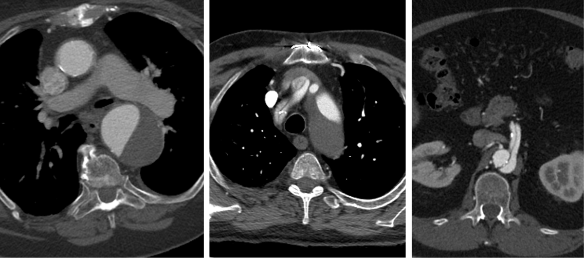
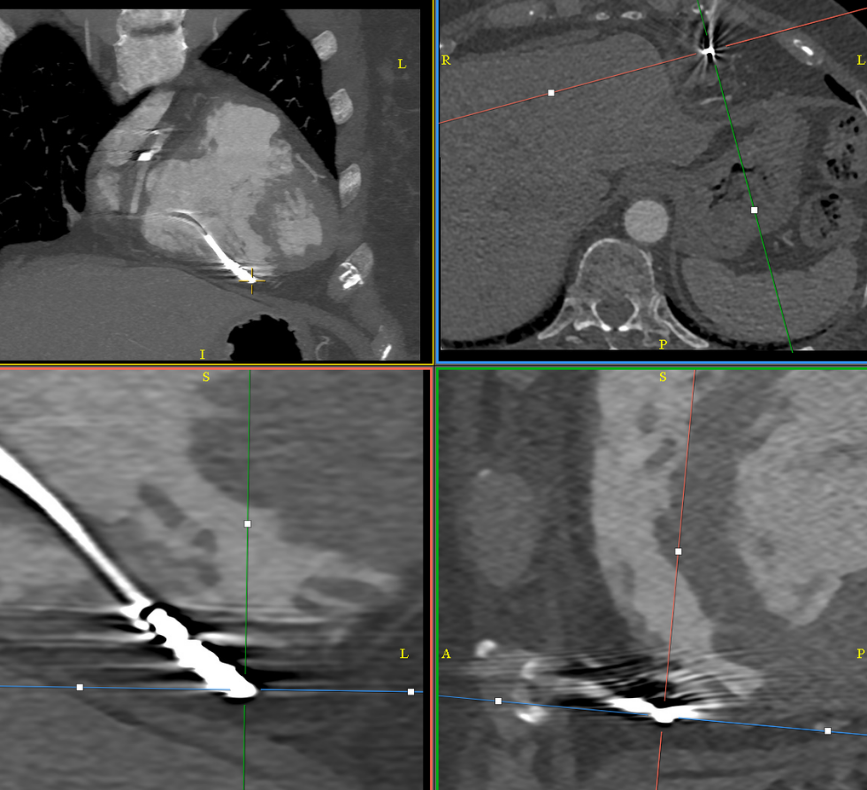
High-risk features should be identified and described to direct management (surgical, endovascular, or hybrid).
Report true / false lumen extension into coronaries, arch vessels, and visceral arteries.
Shared to improve patient safety and healthcare delivery in the provision of radiology services. The circumstances and facts are changed, altered, or deidentified to preserve confidentiality. Privileges have not been waived.
Practical Insights 
High Risk Features:
-
- Hemothorax / hemopericardium.
- End organ infarction.
- Aortic diameter >40mm, false lumen diameter >22mm.
- Entry tear on the lesser curvature of the aorta.
- Refractory pain and/or hypertension.
 Takeaway: Accurate characterization of dissections improves survival.
The Interventional Radiology National Subspecialty Division (NSD) is part of RP’s Clinical Value Team, which works to elevate patient care and enhance value through innovation, collaboration and education. To advance this goal, our radiologists and advanced practice providers are committed to sharing peer learning as valuable reminders and insights about what we encounter in our day-to-day practice. Check back here and on X, LinkedIn and Instagram to see these common cases and our findings.
Visit the Clinical Resources page for more cases and to see what we’ve developed to enhance best practice recommendations, elevate image quality and patient care and update current standards throughout RP’s network of practices, all to deliver excellent radiology services to patients, referring clinicians and client partners.
Radiology Partners, through its owned and affiliated practices, is a leading physician-led and physician-owned technology-enabled radiology practice in the U.S. For the latest news from RP, follow us on X, LinkedIn, Instagram, YouTube and the blog.
 Shared to improve patient safety and healthcare delivery in the provision of radiology services. The circumstances and facts are changed, altered, or deidentified to preserve confidentiality. Privileges have not been waived.
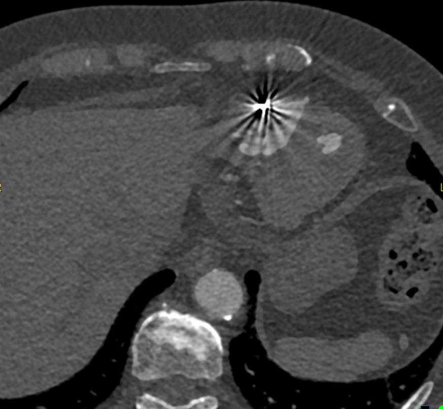
 Shared to improve patient safety and healthcare delivery in the provision of radiology services. The circumstances and facts are changed, altered, or deidentified to preserve confidentiality. Privileges have not been waived.
 Shared to improve patient safety and healthcare delivery in the provision of radiology services. The circumstances and facts are changed, altered, or deidentified to preserve confidentiality. Privileges have not been waived.
 Shared to improve patient safety and healthcare delivery in the provision of radiology services. The circumstances and facts are changed, altered, or deidentified to preserve confidentiality. Privileges have not been waived.
 Shared to improve patient safety and healthcare delivery in the provision of radiology services. The circumstances and facts are changed, altered, or deidentified to preserve confidentiality. Privileges have not been waived.
 Shared to improve patient safety and healthcare delivery in the provision of radiology services. The circumstances and facts are changed, altered, or deidentified to preserve confidentiality. Privileges have not been waived.