Rad to Rad Learning: Pott’s Puffy Tumor

The Radiology Partners (RP) Neuroradiology National Subspecialty Division (NSD) presents our newest Rad to Rad Learning case. 


Peer Learning Opportunitymegaphone icon

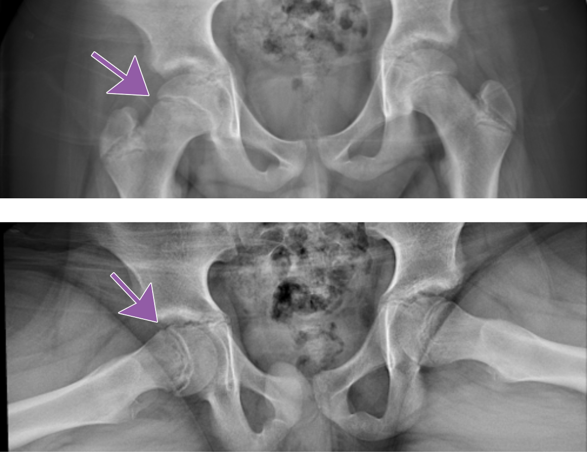
The possibility of intracranial extension of sinus infection results in high morbidity.  

Pott’s Puffy Tumor 

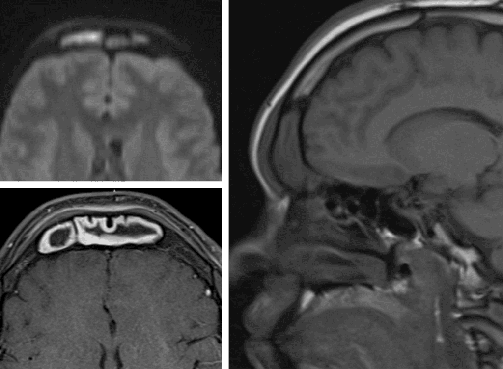
Subperiosteal abscess related to extension of acute frontal sinusitis through the calvarium.

Shared to improve patient safety and healthcare delivery in the provision of radiology services. The circumstances and facts are changed, altered, or deidentified to preserve confidentiality. Privileges have not been waived.


Practical Insights duplicate icon

    • Factors that increase the risk of developing Pott’s include trauma, intranasal cocaine use, methamphetamine use, and craniotomy.

    • Commonly associated intracranial extension needs to be evaluated with an MRI.

    • The overlying calvarium can remain intact.

    • DWI sequence is key for abscess evaluation.

    • More common in adolescents.


Bell iconTakeaway: Prompt identification is required to avoid significant neurologic complications. 

The Neuroradiology National Subspecialty Division (NSD) is part of RP’s Clinical Value Team, whichworks to elevate patient care and enhance value through innovation, collaboration and education. To advance this goal, our radiologists and advanced practice providers are committed to sharing peer learning as valuable reminders and insights about what we encounter in our day-to-day practiceCheck back here and on XLinkedIn and Instagram to see these common cases and our findings.

Visit the Clinical Resources page for more cases and to see what we’ve developed to enhance best practice recommendations, elevate image quality and patient care and update current standards throughout RP’s network of practices, all to deliver excellent radiology services to patients, referring clinicians and client partners.

Radiology Partners, through its owned and affiliated practices, is a leading physician-led and physician-owned technology-enabled radiology practice in the U.S. For the latest news from RP, follow us on XLinkedInInstagramYouTube and the blog.


Rad to Rad Learning: Tension Pneumocephalus

The Radiology Partners (RP) Neuroradiology National Subspecialty Division (NSD) presents our newest Rad to Rad Learning case. 


Peer Learning Opportunitymegaphone icon

Progressive increase in intracranial pressure over time is life threatening.

Tension Pneumocephalus 

a ct scan of a tension pneumocephalus

Note the progressive compression of the frontal lobes by air with widening of interhemispheric spaces. 

Shared to improve patient safety and healthcare delivery in the provision of radiology services. The circumstances and facts are changed, altered, or deidentified to preserve confidentiality. Privileges have not been waived.


Practical Insights duplicate icon

    • Occurs most commonly after SDH evacuation, but can also be seen after skill base or sinonasal surgeries, head trauma, and nitrous oxide anesthesia.

    • Treated with ventriculostomy, craniotomy, and dural defect closure.


Bell iconTakeaway: Symptomatic enlarging pneumocephalus is a neurosurgical emergency.

The Neuroradiology National Subspecialty Division (NSD) is part of RP’s Clinical Value Team, whichworks to elevate patient care and enhance value through innovation, collaboration and education. To advance this goal, our radiologists and advanced practice providers are committed to sharing peer learning as valuable reminders and insights about what we encounter in our day-to-day practiceCheck back here and on XLinkedIn and Instagram to see these common cases and our findings.

Visit the Clinical Resources page for more cases and to see what we’ve developed to enhance best practice recommendations, elevate image quality and patient care and update current standards throughout RP’s network of practices, all to deliver excellent radiology services to patients, referring clinicians and client partners.

Radiology Partners, through its owned and affiliated practices, is a leading physician-led and physician-owned technology-enabled radiology practice in the U.S. For the latest news from RP, follow us on XLinkedInInstagramYouTube and the blog.


Rad to Rad Learning: Ectopic Pregnancy

The Radiology Partners (RP) Body Imaging Subspecialty Division (NSD) presents our newest Rad to Rad Learning case. 


Peer Learning Opportunitymegaphone icon

This condition affects 1-2% of pregnancies. With associated bleeding, 1st trimester pregnancies are at a risk of up to 18%. 

Ectopic Pregnancy93-97% of ectopics are tubal. Tubal ring sign has 95% PPV for ectopic. Visible double decidual sign of the intrauterine sac indicates low probability of ectopic. 

arrows pointing to different types of ectopic pregnancies

Fluid in the endo canal is pseudosac (yellow arrow) vs. Ectopic (purple arrow)

Shared to improve patient safety and healthcare delivery in the provision of radiology services. The circumstances and facts are changed, altered, or deidentified to preserve confidentiality. Privileges have not been waived.


Practical Insights duplicate icon

    • Check for adnexal masses and amount of free fluid in the hepatorenal recess to determine significance.

    • Complex free fluid may represent blood products.

    • Quantitative beta HCG levels are required. Levels >2500 are highly suspicious for occult ectopic.

    • Surgery is recommended for ectopic >3.5cm or if cardiac activity is present.


Bell iconTakeaway: Always report size of ectopic. If >3.5cm, surgery is recommended. 

The Body Imaging Subspecialty Division (NSD) is part of RP’s Clinical Value Team, whichworks to elevate patient care and enhance value through innovation, collaboration and education. To advance this goal, our radiologists and advanced practice providers are committed to sharing peer learning as valuable reminders and insights about what we encounter in our day-to-day practiceCheck back here and on XLinkedIn and Instagram to see these common cases and our findings.

Visit the Clinical Resources page for more cases and to see what we’ve developed to enhance best practice recommendations, elevate image quality and patient care and update current standards throughout RP’s network of practices, all to deliver excellent radiology services to patients, referring clinicians and client partners.

Radiology Partners, through its owned and affiliated practices, is a leading physician-led and physician-owned technology-enabled radiology practice in the U.S. For the latest news from RP, follow us on XLinkedInInstagramYouTube and the blog.


Rad to Rad Learning: Anterior Pneumothorax

The Radiology Partners (RP) Pediatric Radiology National Subspecialty Division (NSD) presents our newest Rad to Rad Learning case.


Peer Learning Opportunitymegaphone icon

This condition is associated with high morbidity but is difficult to see on supine neonates.

Anterior Pneumothorax

Pediatric chest radiograph

Hallmarks: Deep sulcus sign, no lung marking at the edge of right lung, increased sharpness of the cardiomediastinal border, more prominent on expiration.

Pediatric chest radiograph showing anterior pneumothorax with arrow pointing to landmarksShared to improve patient safety and healthcare delivery in the provision of radiology services. The circumstances and facts are changed, altered, or deidentified to preserve confidentiality. Privileges have not been waived.


Practical Insights duplicate icon

    • May be bilateral.

    • Can occur in both term and preterm babies.

    • Compare lung lucency between both sides.

    • Decubitus radiograph can be a helpful tool for confirmation.


Bell iconTakeaway: Even a suspected pneumothorax is a critical result and should be called.

The Pediatric Radiology National Subspecialty Division (NSD) is part of RP’s Clinical Value Team, whichworks to elevate patient care and enhance value through innovation, collaboration and education. To advance this goal, our radiologists and advanced practice providers are committed to sharing peer learning as valuable reminders and insights about what we encounter in our day-to-day practiceCheck back here and on XLinkedIn and Instagram to see these common cases and our findings.

Visit the Clinical Resources page for more cases and to see what we’ve developed to enhance best practice recommendations, elevate image quality and patient care and update current standards throughout RP’s network of practices, all to deliver excellent radiology services to patients, referring clinicians and client partners.

Radiology Partners, through its owned and affiliated practices, is a leading physician-led and physician-owned technology-enabled radiology practice in the U.S. For the latest news from RP, follow us on XLinkedInInstagramYouTube and the blog.


Rad to Rad Learning: Morton’s Neuroma

The Radiology Partners (RP) MSK National Subspecialty Division (NSD) presents our newest Rad to Rad Learning case.


Peer Learning Opportunitymegaphone icon

This finding represents a compressive neuropathy of the interdigital nerve primarily in the 2nd and 3rd intermetatarsal spaces.

Morton’s Neuroma

Moron's Neuroma radiographic imageShared to improve patient safety and healthcare delivery in the provision of radiology services. The circumstances and facts are changed, altered, or deidentified to preserve confidentiality. Privileges have not been waived.


Practical Insights duplicate icon

    • Presents with radiating pain, throbbing, numbness, and burning from the webspace into the toes. (Walking on marbles.)

    • Differential diagnosis: fracture, osteonecrosis, bursitis, synovial cyst.

    • Can have concomitant intermetatarsal bursitis.

    • AKA Intermetatarsal perineural fibrosis.


Bell iconTakeaway: Common cause of forefoot pain – best seen on T1 weighted sequences.

The MSK National Subspecialty Division (NSD) is part of RP’s Clinical Value Team, whichworks to elevate patient care and enhance value through innovation, collaboration and education. To advance this goal, our radiologists and advanced practice providers are committed to sharing peer learning as valuable reminders and insights about what we encounter in our day-to-day practiceCheck back here and on XLinkedIn and Instagram to see these common cases and our findings.

Visit the Clinical Resources page for more cases and to see what we’ve developed to enhance best practice recommendations, elevate image quality and patient care and update current standards throughout RP’s network of practices, all to deliver excellent radiology services to patients, referring clinicians and client partners.

Radiology Partners, through its owned and affiliated practices, is a leading physician-led and physician-owned technology-enabled radiology practice in the U.S. For the latest news from RP, follow us on XLinkedInInstagramYouTube and the blog.


Rad to Rad Learning: Slipped Capital Femoral Epiphysis

The Radiology Partners (RP) Pediatric Radiology National Subspecialty Division (NSD) presents our newest Rad to Rad Learning case.


Peer Learning Opportunitymegaphone icon

Slipped Capital Femoral Epiphysis (SCFE) is a common and urgent diagnosis and needs to be communicated with the referring provider.

Slipped Capital Femoral Epiphysis

radiographic image of the pelvis

Note the medial and posterior displacement of the right femoral head and widened physis. Frog lateral views are generally more sensitive.

radiographic image of the pelvisShared to improve patient safety and healthcare delivery in the provision of radiology services. The circumstances and facts are changed, altered, or deidentified to preserve confidentiality. Privileges have not been waived.


Practical Insights duplicate icon

    • Commonly missed diagnosis that leads to increased complications.

    • Complications include osteonecrosis, chondrolysis, chronic pain, and growth arrest.

    • Having a high index of suspicion is helpful.

    • For at-risk patients, compare the hips on both views.


Bell iconTakeaway: Make sure to communicate positive findings to the referring provider.

The Pediatric Radiology National Subspecialty Division (NSD) is part of RP’s Clinical Value Team, whichworks to elevate patient care and enhance value through innovation, collaboration and education. To advance this goal, our radiologists and advanced practice providers are committed to sharing peer learning as valuable reminders and insights about what we encounter in our day-to-day practiceCheck back here and on XLinkedIn and Instagram to see these common cases and our findings.

Visit the Clinical Resources page for more cases and to see what we’ve developed to enhance best practice recommendations, elevate image quality and patient care and update current standards throughout RP’s network of practices, all to deliver excellent radiology services to patients, referring clinicians and client partners.

Radiology Partners, through its owned and affiliated practices, is a leading physician-led and physician-owned technology-enabled radiology practice in the U.S. For the latest news from RP, follow us on XLinkedInInstagramYouTube and the blog.


Rad to Rad Learning: Mode of Communication

The Radiology Partners (RP) Cardiothoracic Imaging National Subspecialty Division (NSD) presents our newest Rad to Rad Learning case.


Peer Learning Opportunitymegaphone icon

This is a routine outpatient chest x-ray that just showed up on your list at 4:45 pm on a Friday. What is the appropriate management?

Mode of Communication

Chest X-rayShared to improve patient safety and healthcare delivery in the provision of radiology services. The circumstances and facts are changed, altered, or deidentified to preserve confidentiality. Privileges have not been waived.


Practical Insights duplicate icon

    • Call the referring provider right away. Don’t risk the report sitting in their inbox until Monday.

    • We’re all familiar with critical findings that need to be communicated immediately like strokes or PEs, but it is also important to communicate more “routine” findings in a way that would prevent delays in treatment that could cause significant adverse outcomes.


Bell iconTakeaway: Consider potential treatment delays in how you choose to communicate your findings.

The Cardiovascular Imaging National Subspecialty Division (NSD) is part of RP’s Clinical Value Team, whichworks to elevate patient care and enhance value through innovation, collaboration and education. To advance this goal, our radiologists and advanced practice providers are committed to sharing peer learning as valuable reminders and insights about what we encounter in our day-to-day practiceCheck back here and on XLinkedIn and Instagram to see these common cases and our findings.

Visit the Clinical Resources page for more cases and to see what we’ve developed to enhance best practice recommendations, elevate image quality and patient care and update current standards throughout RP’s network of practices, all to deliver excellent radiology services to patients, referring clinicians and client partners.

Radiology Partners, through its owned and affiliated practices, is a leading physician-led and physician-owned technology-enabled radiology practice in the U.S. For the latest news from RP, follow us on XLinkedInInstagramYouTube and the blog.


Rad to Rad Learning: Endoleaks

The Radiology Partners (RP) Interventional Radiology National Subspecialty Division (NSD) presents our newest Rad to Rad Learning case.


Peer Learning Opportunitymegaphone icon

Classification of endoleaks drives critical management and follow-up.

Endoleaks

Type 1: Incomplete endograft seal and contrast tracking around the graft directly into the sac. Type 2: Retrograde flow of one or more branch arteries into the sac.

Shared to improve patient safety and healthcare delivery in the provision of radiology services. The circumstances and facts are changed, altered, or deidentified to preserve confidentiality. Privileges have not been waived.


Practical Insights duplicate icon

    • Type 1:

      • Type 1a = proximal seal.
      • Type 1b = distal seal.
      • Both high-risk, requiring urgent repair!
    • Type 2:

      • Most common.
      • Not urgent, but requires follow-up.
      • For >5mm growth over 6 months, treat with embolization.

Bell iconTakeaway: type 1 endoleak is a critical finding!

The Interventional Radiology National Subspecialty Division (NSD) is part of RP’s Clinical Value Team, whichworks to elevate patient care and enhance value through innovation, collaboration and education. To advance this goal, our radiologists and advanced practice providers are committed to sharing peer learning as valuable reminders and insights about what we encounter in our day-to-day practiceCheck back here and on XLinkedIn and Instagram to see these common cases and our findings.

Visit the Clinical Resources page for more cases and to see what we’ve developed to enhance best practice recommendations, elevate image quality and patient care and update current standards throughout RP’s network of practices, all to deliver excellent radiology services to patients, referring clinicians and client partners.

Radiology Partners, through its owned and affiliated practices, is a leading physician-led and physician-owned technology-enabled radiology practice in the U.S. For the latest news from RP, follow us on XLinkedInInstagramYouTube and the blog.


Rad to Rad Learning: Measuring RV/LV Ratio


The Radiology Partners (RP) Interventional Radiology National Subspecialty Division (NSD) presents our newest Rad to Rad Learning case.


Peer Learning Opportunitymegaphone icon

Patients who have lobar or larger PE and evidence of right heart strain are at increased risk for 30 day in-hospital morbidity and mortality.


Measuring RV/LV Ratio

Measure perpendicular to the long axis of the heart, widest inner wall to inner wall RV diameter and LV diameter.

Measure perpendicular to the long axis of the heart

Right ventricle is 61.5mm. Left ventricle is 19.5mm. RV/LV Ratio is 3.15.

Shared to improve patient safety and healthcare delivery in the provision of radiology services. The circumstances and facts are changed, altered, or deidentified to preserve confidentiality. Privileges have not been waived.


Practical Insights duplicate icon

    • PE with right heart strain is referred to as intermediate risk and warrants assessment.

    • Measure on the slice(s) where each ventricle is widest.

    • An RV / LV ratio ≥ 1.0 = right heart strain

    • The inner wall is the edge of the compacted myocardium.


Bell iconTakeaway: Clot burden must be Central (lobar or main trunk).

The Interventional Radiology National Subspecialty Division (NSD) is part of RP’s Clinical Value Team, whichworks to elevate patient care and enhance value through innovation, collaboration and education. To advance this goal, our radiologists and advanced practice providers are committed to sharing peer learning as valuable reminders and insights about what we encounter in our day-to-day practiceCheck back here and on XLinkedIn and Instagram to see these common cases and our findings.

Visit the Clinical Resources page for more cases and to see what we’ve developed to enhance best practice recommendations, elevate image quality and patient care and update current standards throughout RP’s network of practices, all to deliver excellent radiology services to patients, referring clinicians and client partners.

Radiology Partners, through its owned and affiliated practices, is a leading physician-led and physician-owned technology-enabled radiology practice in the U.S. For the latest news from RP, follow us on XLinkedInInstagramYouTube and the blog.


Rad to Rad Learning: Perched Facets

The Radiology Partners (RP) MSK Musculoskeletal National Subspecialty Division (NSD) presents our newest Rad to Rad Learning case.


Peer Learning Opportunitymegaphone icon

This diagnosis is associated with high risk for spinal cord injury and vertebral artery injury.

[image_with_animation image_url=”9025″ image_size=”large” animation_type=”entrance” animation=”None” animation_movement_type=”transform_y” hover_animation=”none” alignment=”center” border_radius=”none” box_shadow=”none” image_loading=”default” max_width=”100%” max_width_mobile=”default”]

Perched Facets

Malalignment of the facet joints with the inferior articular process of C5 sitting anteriorly “perched” on the superior articular process of C6.

radiographic image of a spinal with malalignment of the facet joints.Shared to improve patient safety and healthcare delivery in the provision of radiology services. The circumstances and facts are changed, altered, or deidentified to preserve confidentiality. Privileges have not been waived.


Practical Insights duplicate icon

    • Mechanism matters: Due to hyperflexion, lateral compression and rotation, most often from automobile accident.

    • Widening of the interspinous distance.

    • Up to 90% of patients with interfacetal dislocation will present with symptoms, and up to 40% can have a complete spinal cord injury.

    • Early diagnosis and treatment can decrease permanent neurologic injury incidence.


Bell iconSearch Pattern alert: Examine facet joints on parasagittal images.

The MSK Musculoskeletal National Subspecialty Division (NSD) is part of RP’s Clinical Value Team, whichworks to elevate patient care and enhance value through innovation, collaboration and education. To advance this goal, our radiologists and advanced practice providers are committed to sharing peer learning as valuable reminders and insights about what we encounter in our day-to-day practiceCheck back here and on XLinkedIn and Instagram to see these common cases and our findings.

Visit the Clinical Resources page for more cases and to see what we’ve developed to enhance best practice recommendations, elevate image quality and patient care and update current standards throughout RP’s network of practices, all to deliver excellent radiology services to patients, referring clinicians and client partners.

Radiology Partners, through its owned and affiliated practices, is a leading physician-led and physician-owned technology-enabled radiology practice in the U.S. For the latest news from RP, follow us on XLinkedInInstagramYouTube and the blog.