A dark blue background with various hexagonal shapes and lines clustered on the left side, representing rad to rad learning, gradually fading into open space on the right.

Rad to Rad Learning: : Epidural Hematoma

The Radiology Partners (RP) Neuroradiology National Subspecialty Division (NSD) presents our newest Rad to Rad Learning case. 


Peer Learning Opportunitymegaphone icon

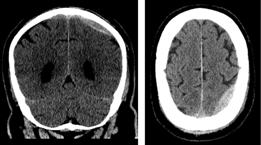
Rapid expansion is common reaching maximum size at 36 hours resulting in neurologic deficits and death in up to 5% due to herniations.

Hyperdense biconvex unilateral extra axial collection adjacent to a skull fracture in 75% of cases.

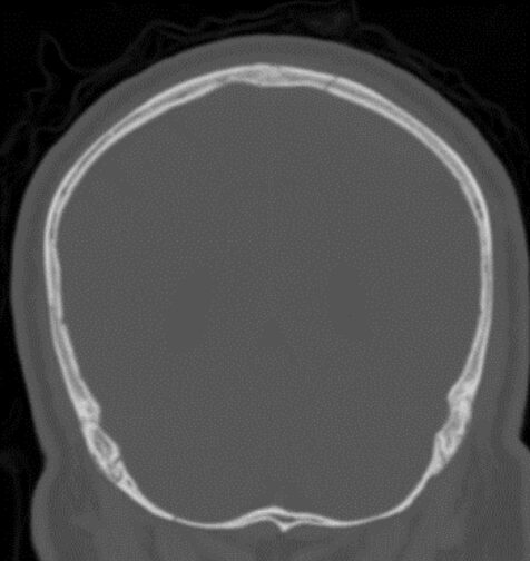
A grayscale scan of a human skull viewed from the front, showing the outline of the skull and cranial cavity with no visible internal detail or abnormalities—a valuable example for Rad to Rad Learning in radiographic interpretation.

Two CT brain scans: the left shows a front-facing (coronal) view, and the right shows a horizontal (axial) view, both revealing brain structures in grayscale with bone shown in white—an ideal example for Rad to Rad Learning.


Practical Insights duplicate icon

    • Common “lucid interval” in up to 50% of cases.
    • Typically associated with arterial laceration, but up to 10% are venous.
    • Consider CTV if collection crosses suture.
    • Bilateral EDH have up to 15-20% mortality rate.

Bell iconTakeaway: Use bone window to look for fracture.

Shared to improve patient safety and healthcare delivery in the provision of radiology services. The circumstances and facts are changed, altered, or deidentified to preserve confidentiality. Privileges have not been waived.The Neuroradiology National Subspecialty Division (NSD) is part of RP’s Clinical Value Team, which works to elevate patient care and enhance value through innovation, collaboration and education. To advance this goal, our radiologists and advanced practice providers are committed to sharing peer learning as valuable reminders and insights about what we encounter in our day-to-day practice. Check back here and on X, LinkedIn and Instagram to see these common cases and our findings.

Visit the Clinical Resources page for more cases and to see what we’ve developed to enhance best practice recommendations, elevate image quality and patient care and update current standards throughout RP’s network of practices, all to deliver excellent radiology services to patients, referring clinicians and client partners.

Radiology Partners, through RP Clinical Services and its managed practices, together with Mosaic Clinical Technologies, Inc., its technology and AI division, is the leading provider of technology-enabled radiology services in the U.S., serving more than 3,400 hospitals and other healthcare facilities with high-quality radiology, technology and artificial intelligence solutions. As a physician-led and physician-owned practice, our mission is to transform radiology by innovating across clinical value, technology, and service, while elevating the role of radiology and radiologists in healthcare. For the latest news from RP, follow us on X, LinkedIn, Instagram, YouTube and the blog.


Two logos on a dark blue background: mosaic CLINICAL TECHNOLOGIES with a star-shaped icon above, and Cognita, an FDA contract partner, with a circular, wave-like icon to the left of the text.

Cognita Imaging Receives $1.29 Million FDA Contract to Test New Approach to Evaluating Generative AI in Radiology

Research will test whether multiple language models can work as a jury to assess AI-generated reports across approximately 1 million patient exams, with radiologists reviewing clinically important discrepancies 

NASHVILLE, Tenn., and PALO ALTO, Calif., (September 16, 2026) — Cognita Imaging, Inc. (Cognita), a wholly owned subsidiary of Mosaic Clinical Technologies, Inc. (Mosaic), has received a $1.29 million research contract from the U.S. Food and Drug Administration (FDA) to test a new way of evaluating AI-generated radiology reports. The contract took effect June 22, 2026, and will run for 18 months. 

The project, titled “Virtual Subject Matter Expert Agents for Radiology Report Evaluation,” focuses on a growing challenge in medical AI, which is how to rigorously evaluate systems that generate complete radiology reports. Traditional reader studies are essential, but expert review takes time and is difficult to extend to the number and variety of cases needed to understand how a model may behave in day-to-day practice. 

Under the contract, Cognita will develop and validate a framework that uses several large language models (LLMs) to evaluate human-drafted and AI-generated radiology reports. The work will be led by Akshay Chaudhari, Ph.D., Cognita co-founder, associate professor of radiology and biomedical data science at Stanford University and principal investigator on the contract. Louis Blankemeier, Ph.D., co-founder and chief executive officer of Cognita, is the co-investigator. 

“Once an AI system starts writing the entire report, the evaluation problem changes,” said Chaudhari. “A few hundred cases may tell you whether a model works in a narrow setting. They do not show every way it can fail in practice. We want to test whether a group of language models, with radiologists reviewing the difficult cases, can give us a clearer and more repeatable picture at real-world scale.” 

Cognita calls the method “LLMs-as-a-jury.” Rather than asking one language model to grade another model’s report, the framework will compare the judgments of several models. The goal is to make the evaluation more consistent, reduce dependence on the tendencies of any one model and identify where automated review can meaningfully extend expert evaluation. 

After building and testing the framework, Cognita will apply it to approximately 1 million patient exams from a large and diverse U.S. cohort. Researchers will examine performance across different patient groups, care settings, imaging equipment and diseases, including uncommon findings that may appear too rarely to be studied well in a smaller dataset. The team will also recreate smaller validation cohorts from the larger dataset to determine what information may be missed when a study includes fewer cases. 

Radiologists will review clinically important disagreements and determine whether the problem came from the AI-generated report, the language-model jury or the original radiologist’s report. Cognita will use those findings to improve the framework and study how automated review and physician expertise can work together in both premarket validation and post-market monitoring. 

“Radiologists know the edge cases matter,” said Nina Kottler, M.D., chief medical AI officer at Mosaic Clinical Technologies. “A model can look strong in a carefully selected study and still struggle at another hospital, on different equipment or with a rare presentation. This work lets us look for those differences at a scale that would be very difficult to reach through reader studies alone, while keeping radiologists involved where their judgment matters most.” 

The research builds on Cognita’s prior work in radiology-specific language and vision-language models. That work includes GREEN, an open-source tool that compares reference and AI-generated radiology reports and identifies clinically meaningful differences. Cognita will also draw on clinical expertise, technology infrastructure and real-world radiology workflows from the broader Mosaic and Radiology Partners Clinical Services’ ecosystem. 

During the contract, Cognita will provide FDA with software code and practical guidance for building LLM juries, analyses comparing large and small validation cohorts, radiologist-reviewed discrepancy studies, and reports on the project’s findings and limitations. The contract was awarded through FDA’s Broad Agency Announcement program for advanced research and development in regulatory science. 

About Cognita Imaging, Inc. 

Cognita Imaging, Inc. develops vision-language models and other generative AI capabilities for radiology. Its work is focused on helping radiologists deliver accurate, efficient care and expanding access to high-quality health care. Cognita seeks to elevate the performance of all radiologists to the level of the best expert subspecialists and, longer term, to empower radiologists with digital imaging information not easily perceptible to the human eye. Cognita is a wholly owned subsidiary of Mosaic Clinical Technologies, Inc. Learn more at cognita.ai. 

About Mosaic Clinical Technologies, Inc. 

Mosaic Clinical Technologies, Inc., a wholly owned subsidiary of Radiology Partners, Inc., develops technology for medical imaging. Its MosaicOS™ platform brings diagnostic tools, AI and radiology workflows together in a cloud-native operating system designed to support radiologists and the organizations they serve. Learn more at MosaicClinical.ai. 

About Radiology Partners, Inc. 

Radiology Partners, Inc., through RP Clinical Services and its managed practices, together with Mosaic Clinical Technologies, Inc., provides radiology services and technology to more than 3,400 hospitals and other health care facilities. Its network includes more than 4,000 radiologists who interpret more than 55 million imaging studies each year. Learn more at radpartners.com. 

Media Contact
Ink House
Cognita@inkhouse.com


A dark blue background with various hexagonal shapes and lines clustered on the left side, representing rad to rad learning, gradually fading into open space on the right.

Rad to Rad Learning: Cerebral Amyloid Angiopathy

The Radiology Partners (RP) Neuroradiology National Subspecialty Division (NSD) presents our newest Rad to Rad Learning case. 


Peer Learning Opportunitymegaphone icon

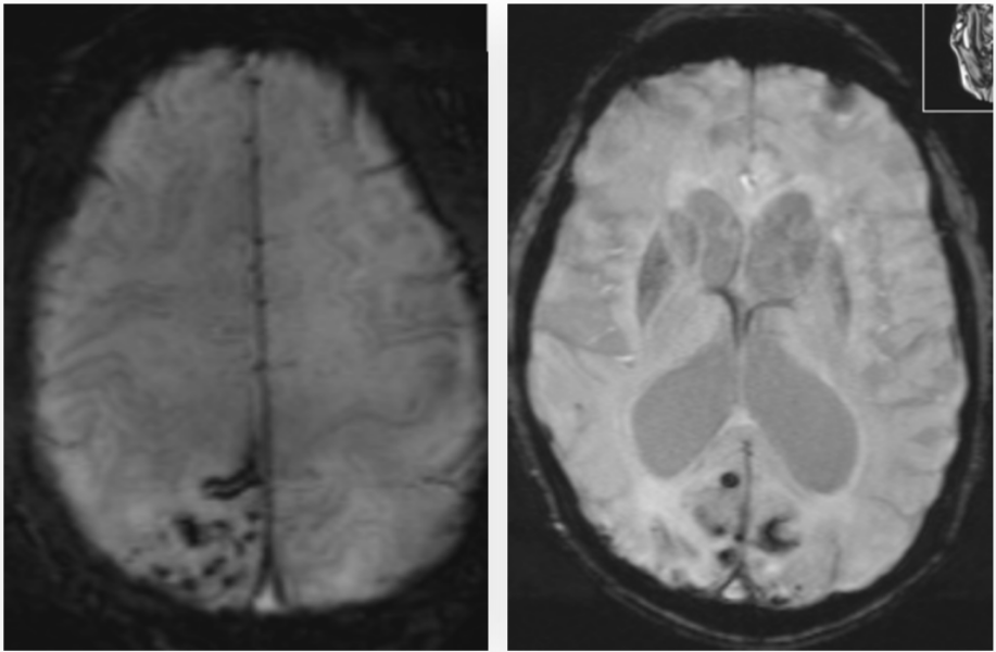
Early imaging-based diagnosis is critical, as hemorrhagic complications can be mitigated by avoiding certain therapies.

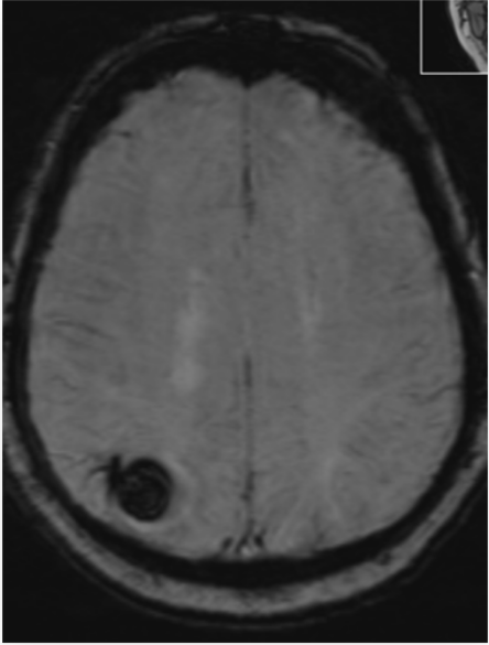
Superficial siderosis, sequalae of bilateral microhemorrhages and large parenchymal hemorrhage.

Axial MRI brain scan showing a circular, dark lesion near the left posterior region, possibly indicating a mass or abnormality—an ideal case for Rad to Rad Learning and collaborative diagnostic review.


Practical Insights duplicate icon

    • Causes up to 1/5 of primary intracranial hemorrhage in patients >60 years.
    • Boston criteria are used for diagnosis without biopsy using presence of multiple asymmetric lobar microhemorrhages, cortical superficial siderosis, and lobar ICH.
    • Other markers are multiple dilated perivascular spaces and white matter T2 hyperintensities (70%).
    • May show sulcal or leptomeningeal enhancement or focal nonhemorrhagic mass (amyloidoma) extending to the ventricular wall in younger patients.

Bell iconTakeaway: Suspect CAA in normotensive patients with multiple types of non-traumatic hemorrhages.

Shared to improve patient safety and healthcare delivery in the provision of radiology services. The circumstances and facts are changed, altered, or deidentified to preserve confidentiality. Privileges have not been waived.The Neuroradiology National Subspecialty Division (NSD) is part of RP’s Clinical Value Team, which works to elevate patient care and enhance value through innovation, collaboration and education. To advance this goal, our radiologists and advanced practice providers are committed to sharing peer learning as valuable reminders and insights about what we encounter in our day-to-day practice. Check back here and on X, LinkedIn and Instagram to see these common cases and our findings.

Visit the Clinical Resources page for more cases and to see what we’ve developed to enhance best practice recommendations, elevate image quality and patient care and update current standards throughout RP’s network of practices, all to deliver excellent radiology services to patients, referring clinicians and client partners.

Radiology Partners, through RP Clinical Services and its managed practices, together with Mosaic Clinical Technologies, Inc., its technology and AI division, is the leading provider of technology-enabled radiology services in the U.S., serving more than 3,400 hospitals and other healthcare facilities with high-quality radiology, technology and artificial intelligence solutions. As a physician-led and physician-owned practice, our mission is to transform radiology by innovating across clinical value, technology, and service, while elevating the role of radiology and radiologists in healthcare. For the latest news from RP, follow us on X, LinkedIn, Instagram, YouTube and the blog.


A dark blue background with various hexagonal shapes and lines clustered on the left side, representing rad to rad learning, gradually fading into open space on the right.

Rad to Rad Learning: Osteomyelitis

The Radiology Partners (RP) MSK National Subspecialty Division (NSD) presents our newest Rad to Rad Learning case. 


Peer Learning Opportunitymegaphone icon

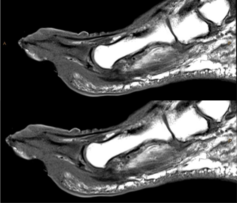
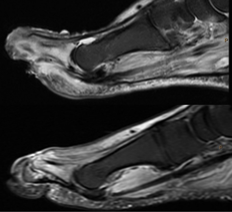
T1 marrow replacement is not necessary to diagnose osteomyelitis in the presence of subjacent ulcers.

T1 signal is darker than muscle (marrow replacement).

T1 signal is darker than muscle (marrow replacement).

T2 edema signal.

T2 edema signal.


Practical Insights duplicate icon

    • Bone marrow edema signal with T1 marrow replacement is consistent with osteomyelitis.  

    • Bone marrow edema signal without T1 marrow replacement with adjacent ulcer, abscess, or sinus tract indicates “high likelihood of osteomyelitis”.


Bell iconTakeaway: Osteo – likely no contrast needed.

Shared to improve patient safety and healthcare delivery in the provision of radiology services. The circumstances and facts are changed, altered, or deidentified to preserve confidentiality. Privileges have not been waived.The MSK National Subspecialty Division (NSD) is part of RP’s Clinical Value Team, which works to elevate patient care and enhance value through innovation, collaboration and education. To advance this goal, our radiologists and advanced practice providers are committed to sharing peer learning as valuable reminders and insights about what we encounter in our day-to-day practice. Check back here and on X, LinkedIn and Instagram to see these common cases and our findings.

Visit the Clinical Resources page for more cases and to see what we’ve developed to enhance best practice recommendations, elevate image quality and patient care and update current standards throughout RP’s network of practices, all to deliver excellent radiology services to patients, referring clinicians and client partners.

Radiology Partners, through RP Clinical Services and its managed practices, together with Mosaic Clinical Technologies, Inc., its technology and AI division, is the leading provider of technology-enabled radiology services in the U.S., serving more than 3,400 hospitals and other healthcare facilities with high-quality radiology, technology and artificial intelligence solutions. As a physician-led and physician-owned practice, our mission is to transform radiology by innovating across clinical value, technology, and service, while elevating the role of radiology and radiologists in healthcare. For the latest news from RP, follow us on X, LinkedIn, Instagram, YouTube and the blog.


A dark blue background with various hexagonal shapes and lines clustered on the left side, representing rad to rad learning, gradually fading into open space on the right.

Rad to Rad Learning: VQ Scan

The Radiology Partners (RP) NMMI Radiology National Subspecialty Division (NSD) presents our newest Rad to Rad Learning case. 


Peer Learning Opportunitymegaphone icon

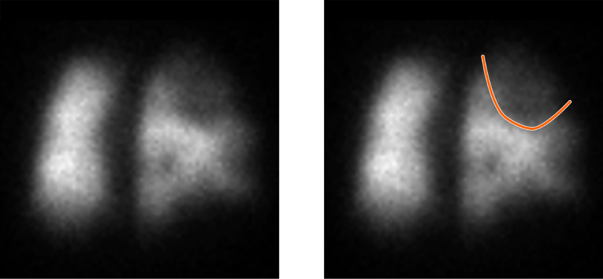
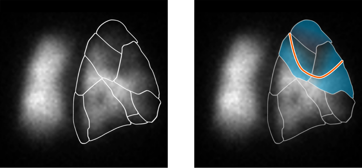
PIOPED criteria relies on the size of segmental perfusion defects to determine the probability of pulmonary embolism.

Use the lung segmental map as a guide for determining defect size. This “single” defect is actually three segmental defects, one large and two medium sized.

Two blurry grayscale images side by side show vague shapes. The right image has an orange curved line drawn over its upper right area, while the left image has no markings, illustrating a typical scenario in Rad to Rad Learning visualizations.

Two grayscale images of a lung scan: the left image shows lung regions outlined in white, while the right highlights the upper-right lung region in blue with an orange border, demonstrating detailed focus on that area as part of Rad to Rad Learning.


Practical Insights duplicate icon

    • Small defect <25% of a lung segment.

    • Medium defect >25& to <75% of a lung segment.

    • Large defect >75% of a lung segment.

    • One visible defect may involve two or more segments.

    • Correlate with chest x-ray within 24 hours.


Bell iconTakeaway: The number and size of segmental defects matters in VQ scans.

Shared to improve patient safety and healthcare delivery in the provision of radiology services. The circumstances and facts are changed, altered, or deidentified to preserve confidentiality. Privileges have not been waived.The NMMI Radiology National Subspecialty Division (NSD) is part of RP’s Clinical Value Team, which works to elevate patient care and enhance value through innovation, collaboration and education. To advance this goal, our radiologists and advanced practice providers are committed to sharing peer learning as valuable reminders and insights about what we encounter in our day-to-day practice. Check back here and on X, LinkedIn and Instagram to see these common cases and our findings.

Visit the Clinical Resources page for more cases and to see what we’ve developed to enhance best practice recommendations, elevate image quality and patient care and update current standards throughout RP’s network of practices, all to deliver excellent radiology services to patients, referring clinicians and client partners.

Radiology Partners, through RP Clinical Services and its managed practices, together with Mosaic Clinical Technologies, Inc., its technology and AI division, is the leading provider of technology-enabled radiology services in the U.S., serving more than 3,400 hospitals and other healthcare facilities with high-quality radiology, technology and artificial intelligence solutions. As a physician-led and physician-owned practice, our mission is to transform radiology by innovating across clinical value, technology, and service, while elevating the role of radiology and radiologists in healthcare. For the latest news from RP, follow us on X, LinkedIn, Instagram, YouTube and the blog.


Blurred image with cool blue and white tones, evoking the visual harmony of Anthem Blue Cross and Blue Shield of Georgia and Northside Radiology Associates Announce Multi-Year Agreement. The abstract, unfocused appearance features no distinct shapes or objects, creating a soft and dreamy atmosphere that mirrors the seamless collaboration between these trusted partners.

Radiology Partners to Acquire Everlight Radiology, Creating a Global Leader in Teleradiology

Combination will extend 24/7/365 subspecialty coverage for hospitals, health systems and imaging providers across the United Kingdom, Ireland, Australia, New Zealand and South Africa 

(NASHVILLE, Tenn., and LONDON) – August 25, 2026  – Radiology Partners, Inc. (“RP”), the leading provider of technology and AI-enabled radiology services, today announced that it has entered into a definitive agreement to acquire Everlight Radiology (“Everlight”), a leading international teleradiology provider serving hospitals, health systems and imaging centers across the United Kingdom, Ireland, Australia, New Zealand and South Africa. The transaction is subject to customary regulatory approvals and closing conditions.

The combination brings together capabilities from Everlight’s international radiologist network and RP Clinical Services’ vRad unit, the largest teleradiology platform in the U.S., to form a leader in global teleradiology. Each organization will continue to serve the markets and clients it serves today, with radiologists reading where they are licensed and credentialed. The two will share technology and clinical standards so that clients in every market benefit from tools and quality standards proven across the combined business.

The shortage of radiologists is an acute and global one. For example, in the United Kingdom, according to the Royal College of Radiologists, demand for CT and MRI imaging grew by 9% in 2024-25 while the radiologist workforce grew by 4.7%, a persistent trend that has led to a shortfall of thousands of radiologists necessary to meet the growing demand. In Australia, according to government figures, the number of diagnostic imaging services under the country’s Medicare system rose by around 40% over the past decade, with advanced imaging growing at an almost 2x faster rate than radiologist workforce supply. Health systems across North America, Europe, Asia and Africa are managing versions of the same shortfall, felt most acutely overnight, on weekends and holidays, when patients still need urgent studies reported and local coverage is at its thinnest.

Teleradiology has become a key approach to closing coverage gaps as it allows for load balancing, subspecialty balancing, technology standardization and the innovative application of artificial intelligence tools to extend local market capacity where supply is limited. Both organizations have heavily invested in technology for years – Everlight through its in-house technology team and clinical AI partnerships, and Radiology Partners through vRad, which has been developing clinical AI models since 2015; Mosaic Clinical Technologies, Inc., its technology and AI division, whose MosaicOS™ platform integrates advanced vision language AI models, AI-enabled reporting and workflow tools into a single system, and Cognita AI its advanced artificial intelligence lab. Together, they intend to accelerate technology advancements that improve radiologist productivity and capacity, while simultaneously improving quality.

Mosaic Drafting, which applies proprietary AI models to interpret imaging exams and create draft reports for radiologist review and sign-off, all fully integrated into advanced AI reporting tools. Subject to the required regulatory clearances in each market, Radiology Partners intends to make Mosaic Drafting available to Everlight radiologists.

Founded in 2006, Everlight operates a “follow-the-sun” model that draws on a network of radiologists working across multiple countries and time zones, so that an urgent study generated overnight in one market can be reported by a subspecialty-trained radiologist in another market during their daytime hours. Everlight’s network comprises more than 800 radiologists working in over 40 countries, reporting over two-and-a-half million exams a year for 340-plus client organizations. The model is designed so that reporting capacity is available continuously rather than concentrated in a single national workforce.

Radiology Partners, through RP Clinical Services, serves more than 3,400 hospitals and other healthcare facilities in the U.S. and cares for tens of millions of unique patients a year. Its national teleradiology practice, vRad, has operated since 2001 and has cumulatively served nearly 120 million unique patients, with a proprietary technology platform supported by 25 patents and more than 25 clinical AI models. As a physician-led and physician-owned practice, RP has built its technology to empower its radiologists to deliver exceptional quality while delivering much needed capacity to the clients we serve.

“Everlight and Radiology Partners have each built something rare in radiology. Everlight has an unmatched global network of subspecialty radiologists, built over 20 years. RP has likewise spent many years in teleradiology and built the clinical infrastructure and technology that supports radiologists at scale,” said Rich Whitney, Chairman and Chief Executive Officer of Radiology Partners. “Together, we can bring the best of what each has built to a growing number of patients and hospitals, delivering more advanced technology, deeper subspecialty support and higher standards of care in every market in which we operate.”

For Everlight’s clients, day-to-day relationships, contacts and clinical workflows are unchanged, and the business will continue to be led by its current management team.

“We are thrilled to be joining Radiology Partners, a move that will provide our radiologists and our clients access to additional clinical depth, technology and investment at a scale that would take years to build alone,” said Rob Anderson, Global Chief Executive Officer of Everlight Radiology. “What will not change is what our clients value most: the same teams, the same standards and the same commitment to reporting every study quickly and accurately.”

Everlight has been majority owned since 2021 by Livingbridge, the UK-based private equity firm. The transaction is expected to close following the required regulatory clearances, after which Everlight will operate as part of Radiology Partners’ global teleradiology business. Rothschild & Co. and Barclays are acting as financial advisors to Radiology Partners, Jones Day and Kirkland & Ellis are serving as its legal counsel.

About Radiology Partners
Radiology Partners, Inc., through RP Clinical Services and its managed practices, together with Mosaic Clinical Technologies, Inc., its technology and AI division, is the leading provider of technology-enabled radiology services in the U.S., serving more than 3,400 hospitals and other healthcare facilities with high-quality radiology, technology and artificial intelligence solutions. As a physician-led and physician-owned practice, our mission is to transform radiology by innovating across clinical value, technology and service, while elevating the role of radiology and radiologists in healthcare. Using a proven healthcare services model, RP Clinical Services provides consistent, high-quality care to patients, while delivering enhanced value to the hospitals, clinics, imaging centers and referring physicians we serve. Within RP Clinical Services, vRad (Virtual Radiologic) is a pioneer of teleradiology, expanding access to diagnostic imaging for millions of patients each year through a practice of more than 500 U.S. board-certified radiologists, the majority of whom are subspecialty trained. Mosaic Clinical Technologies, Inc., powers that work through MosaicOS™, a proprietary imaging intelligence platform that integrates diagnostic technologies, large vision language models, AI-powered tools and smart workflows into a single scalable solution, supporting RP Clinical Services’ national network of practices and commercial partners across the imaging landscape.

Learn more about Radiology Partners at radpartners.com and connect with us on LinkedIn, X, Instagram and YouTube.

Learn more about vRad at vrad.com.

Learn more about Mosaic Clinical Technologies, Inc. at mosaicclinical.ai and connect with us on LinkedIn and YouTube.

About Everlight Radiology
For 20 years, Everlight Radiology has been a lifeline to hospitals, private imaging clinics and corporate radiology providers across the UK, Ireland, Australia, New Zealand and more recently South Africa. A network of over 800+ Consultant Radiologists deliver over 2.5 million urgent, sub-specialist, and routine radiology reports to healthcare providers and hospitals around the globe, fulfilling the mission to enable timely and effective treatment, unlocking the best care for patients and their families by providing worldwide access to specialist radiologists.

Find out more about Everlight Radiology at everlightradiology.com.

Notes to Editors

  1. Royal College of Radiologists; Clinical Radiology Workforce Census 2025, published June 2026. Link.
  2. Australian Institute of Health and Welfare; Pathology, Imaging and Other Diagnostic Services, updated July 2026. Link.

Media Contacts
Sheila Biggs
sbiggs@jarrardinc.com


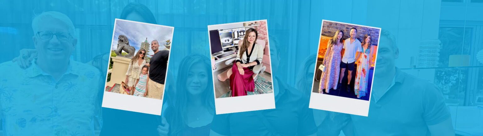
Four people stand together behind three polaroid photos featuring various group and individual RP scenes. The background is overlaid with a blue tint, partially obscuring the group.

Why RP? A Q&A with Dr. Carla Ramas, Body Radiologist

Dr. Carla Ramas reflects on her unconventional path to radiology and how the specialty has allowed her to build a fulfilling career at Radiology Partners (RP) with balance outside of medicine. 

Dr. Carla Ramas is a remote body radiologist based in South Florida. She joined Matrix at RP, the internal remote radiology practice of RP, in December 2020. Outside of work, Dr. Ramas enjoys the outdoors, visiting the beach, traveling, photography and spending time with her family.  

We talked to Dr. Ramas about why she chose Matrix, her passion for supporting the radiologist experience, and her excitement about shaping the future of radiology through MosaicOS™.  


Tell us about why you chose radiology.
Interestingly enough, radiology was not on my radar when I started medical school about 20 years ago. I went into medicine thinking I would become a surgeon. Then, during fourth year of medical school, I did a radiology elective and was surprised by how much I loved it, but thought it was too late to change course because I was already headed into surgery. During my general surgery internship though, I realized surgery was not the right fit for me, and I started thinking, “This does not have to be it. Maybe there is a way to switch into radiology.” I sent out my CV and emailed radiology program directors to explain my situation, but there were no open spots. Then, during my last month of internship, a radiology program director reached out and said someone in his program was leaving radiology to switch to surgery. I interviewed, and he offered me the spot right then. I did not waste any time. My general surgery internship counted as my preliminary year, and I went straight into radiology. It was honestly one of the best decisions I have ever made. I love radiology, I love what I do, and it has allowed me to build a fulfilling career while still having a personal life outside of medicine. To me, it is the best of both worlds. 

What drew you to your subspecialty?
I felt like body imaging was an extension of what interested me in general surgery. That’s the part of the anatomy that I enjoy: the body. During my radiology residency, one of my mentors was a body imaging specialist, and I admired him and learned so much from him. I think having a mentor really helps shape what your interests turn out to be. 

How did you connect with RP?
I knew early on that I wanted to do teleradiology. I wanted my own life outside of medicine, which was an important lesson I learned during my surgery internship, so I looked into teleradiology right out of fellowship. When I started, I did mostly deep overnights, which was the biggest need in teleradiology at that time; there was little daytime work. I started getting burnt out with the deep overnight schedule and wanted to switch to days. One of my friends worked at Matrix and said they had daytime positions available, and that’s what prompted me to make the move. I interviewed with Matrix, and I liked everything I heard. That was over five years ago.  

What do you like about Matrix?
The best thing about Matrix is the people I work with. My co-radiologists, the support team and the leadership are amazing. I love coming to work and hopping onto Teams where we have our group chat, sharing interesting cases. You never feel alone or isolated, which was one of the biggest concerns I had about doing teleradiology. 

As a radiologist at RP, you’re a user of MosaicOS. What does that mean for you as a practicing radiologist to have the ability to help shape the tools you will be using to try to keep up with the future? 
I think it is wonderful. I like being part of something that is helping move radiology forward. AI is going to be an important part of our future, and I think radiologists have an opportunity to help shape how it is used in a way that truly supports our work and improves patient care. We have seen major changes in the field before. When I started residency, people were still using hard films and transcriptionists, and now digital imaging and voice recognition are simply part of how we practice. Mosaic feels like the next evolution. What is exciting to me is that radiologists are not just using Mosaic; we are influencing how it works. That means the tools can be designed around the realities of our day-to-day work, helping us become more efficient, more focused and better able to deliver high-quality radiology services for the patients and referring providers who depend on us. It is a powerful tool, and I look forward to seeing how it continues to evolve. 

What excites you about utilizing Mosaic in your daily practice?
I think it is incredibly promising because it is already making a difference in practical, everyday ways. For example, if I am dictating a body case and identify an incidental liver lesion, I used to have to go to a separate screen, find the appropriate American College of Radiology (ACR) guideline and copy it into the report. With Mosaic, that step is built into the workflow. I describe the finding, include the measurement, and when I process the report, the appropriate guideline is automatically added into the impression. That may seem like a small thing, but it takes a significant mental load off the radiologist. It helps reduce extra steps and saves me time. It frees up my mental energy so I can stay focused on the case in front of me. That is what excites me most: seeing technology support the way we actually practice and help us deliver clear, consistent, high-quality reports for patients and referring providers. 

What would you tell a radiologist who is considering joining Radiology Partners?
If somebody is considering joining RP, I would encourage them to look into it, talk to our radiologists and see the practice for themselves. One of the greatest strengths of RP is the depth and breadth of subspecialty expertise across the practice. As a body imager, there are certain cases I do not routinely read, like MSK MRI. At RP, I have colleagues on shift who are specialty trained in those areas. The same is true in reverse: if an MSK radiologist has a body MRI, I can read that case. That kind of support matters because it helps ensure each study is interpreted by the radiologist best suited for that case. To me, that is ultimately about delivering the best radiology care for patients. We can provide the most accurate, high-quality reports because we have a large, collaborative group with a wide range of specialties. 

What would you say to encourage a young medical student to go into radiology?
I think it’s important to shadow a radiologist. Radiology is one of those behind-the-scenes specialties where a lot of lay people don’t even know about us. They think the ER doctor is the one reading their images. But we are the doctor’s doctor. We’re the ones they go to when they have questions about the studies. That’s an invaluable role. So for the right person and the right personality, I think radiology is one of the best specialties you can go into, and it allows you to enjoy medicine and have a personal life. 

What are those hobbies and interests that you get to enjoy?
I live in South Florida, so I love the outdoors, the beach/ocean. I like fashion, photography and good food, and I like to travel. I’ve always had dogs – bull terriers – and I love being at home and being able to spend time with them. They’re great assistants, just sitting in the back snoring while I’m reading cases. I have a 7-year-old daughter, and we love going to the pool, beach and parks. Orlando, the Florida West coast and the Florida Keys are close by, so even staying within Florida, there’s a lot for us to do, and we always try to do a lot with her. We’re always outdoors, trying to enjoy the weather. 

Dr. Carla Ramas earned her medical degree from SUNY Stony Brook School of Medicine; completed her general surgery internship at NYU, her radiology residency at Winthrop University Hospital, and her body imaging fellowship at Stanford University. 

 Radiology Partners, Inc., through RP Clinical Services and its managed practices, together with Mosaic Clinical Technologies, Inc., its technology and AI division, is the leading provider of technology-enabled radiology services in the U.S., serving more than 3,400 hospitals and other healthcare facilities with high-quality radiology, technology and artificial intelligence solutions. As a physician-led and physician-owned practice, our mission is to transform radiology by innovating across clinical value, technology, and service, while elevating the role of radiology and radiologists in healthcare. For the latest news from RP, follow along on our blog and on X, LinkedIn, Instagram and YouTube. Interested in learning about career opportunities? Visit our careers page.    


A dark blue background with various hexagonal shapes and lines clustered on the left side, representing rad to rad learning, gradually fading into open space on the right.

Rad to Rad Learning: Failed Fundoplication

The Radiology Partners (RP) Body Imaging National Subspecialty Division (NSD) presents our newest Rad to Rad Learning case. 


Peer Learning Opportunitymegaphone icon

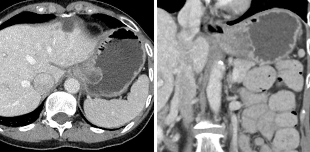
Your next case is this abdominal CT without history or priors. What is the diagnosis? Misdiagnosis may lead to ulceration, necrosis, perforation, and/or bleeding.

Patient has a recent history of fundoplication with gastric wrap around.

CT scans showing a large mass in the upper abdomen, likely originating from the spleen, with clear borders and different densities compared to surrounding tissues. One image is axial, the other coronal—ideal for Rad to Rad Learning and collaborative case discussions.


Practical Insights duplicate icon

    • Fundoplication has a 5 year follow-up failure rate of 4-14&, with 2.8% requiring additional surgery.

    • GERD, pain, and difficulty swallowing are common presenting symptoms.

    • Hx of surgery is key to differentiate from GIST, adenocarcinoma, esophageal varices, gastric wall hematoma or hemorrhage.

    • If there is obstruction, follow up surgery is likely required.


Bell iconTakeaway: History is crucial for narrowing the differential.

Shared to improve patient safety and healthcare delivery in the provision of radiology services. The circumstances and facts are changed, altered, or deidentified to preserve confidentiality. Privileges have not been waived.The Body Imaging National Subspecialty Division (NSD) is part of RP’s Clinical Value Team, which works to elevate patient care and enhance value through innovation, collaboration and education. To advance this goal, our radiologists and advanced practice providers are committed to sharing peer learning as valuable reminders and insights about what we encounter in our day-to-day practice. Check back here and on X, LinkedIn and Instagram to see these common cases and our findings.

Visit the Clinical Resources page for more cases and to see what we’ve developed to enhance best practice recommendations, elevate image quality and patient care and update current standards throughout RP’s network of practices, all to deliver excellent radiology services to patients, referring clinicians and client partners.

Radiology Partners, through RP Clinical Services and its managed practices, together with Mosaic Clinical Technologies, Inc., its technology and AI division, is the leading provider of technology-enabled radiology services in the U.S., serving more than 3,400 hospitals and other healthcare facilities with high-quality radiology, technology and artificial intelligence solutions. As a physician-led and physician-owned practice, our mission is to transform radiology by innovating across clinical value, technology, and service, while elevating the role of radiology and radiologists in healthcare. For the latest news from RP, follow us on X, LinkedIn, Instagram, YouTube and the blog.


A dark blue background with various hexagonal shapes and lines clustered on the left side, representing rad to rad learning, gradually fading into open space on the right.

Rad to Rad Learning: Rapid Gastric Emptying

The Radiology Partners (RP) NMMI Radiology National Subspecialty Division (NSD) presents our newest Rad to Rad Learning case. 


Peer Learning Opportunitymegaphone icon

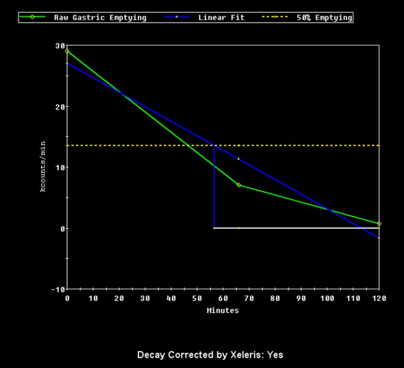
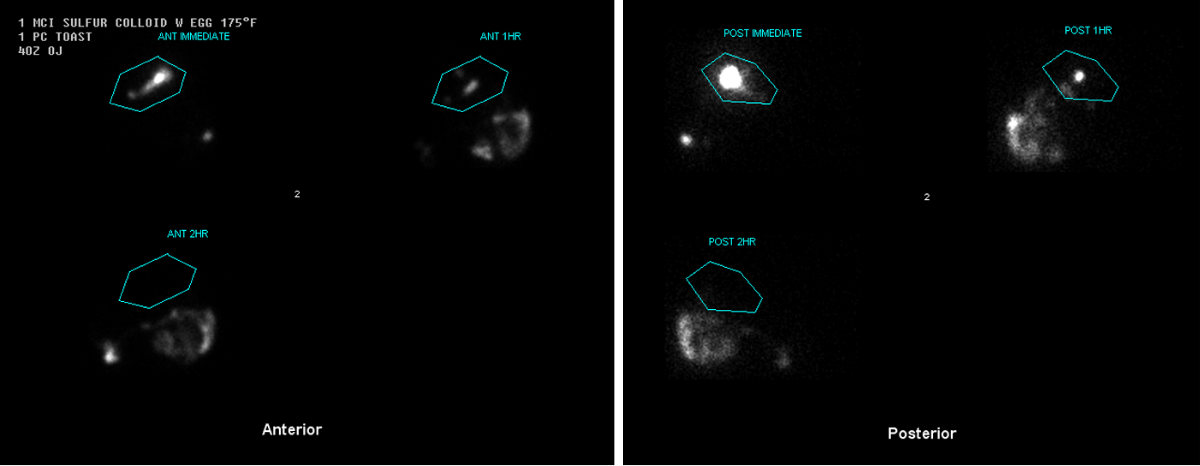
Rapid gastric emptying and gastroparesis can both present with similar clinical symptoms. Gastric emptying examinations can definitively distinguish between the two etiologies.

 1 hour: 24% retention (normal: 30-90%)

This graph displays the change in gastric activity over time at hourly intervals.

A graph showing gastric emptying over 120 minutes with raw and linear fit lines, and a dashed line for 50% emptying. The x-axis is time in minutes; the y-axis is counts per minute. Text below states Decay Corrected by Xeleris: Yes..


Nuclear medicine scan showing anterior and posterior views with highlighted regions labeled ANT MEDLINE, ANT HR, ANT 3HR, POST MEDLINE, POST HR, and POST 3HR in blue outlines.


Practical Insights duplicate icon

    • Seen frequently in patients post gastric surgery, vagotomy, and early diabetes.

    • Diagnostic criteria: <30% retention at 60 minutes.

    • This diagnostic criteria only applies to the standard solid-phase meal, and a combination of anterior and posterior imaging.

    • This examination can be terminated at the 1 hour time point if <30% retention is seen at 60 minutes.


Bell iconTakeaway: Obtaining a detailed patient history is key to an accurate diagnosis.

Shared to improve patient safety and healthcare delivery in the provision of radiology services. The circumstances and facts are changed, altered, or deidentified to preserve confidentiality. Privileges have not been waived.The NMMI Radiology National Subspecialty Division (NSD) is part of RP’s Clinical Value Team, which works to elevate patient care and enhance value through innovation, collaboration and education. To advance this goal, our radiologists and advanced practice providers are committed to sharing peer learning as valuable reminders and insights about what we encounter in our day-to-day practice. Check back here and on X, LinkedIn and Instagram to see these common cases and our findings.

Visit the Clinical Resources page for more cases and to see what we’ve developed to enhance best practice recommendations, elevate image quality and patient care and update current standards throughout RP’s network of practices, all to deliver excellent radiology services to patients, referring clinicians and client partners.

Radiology Partners, through RP Clinical Services and its managed practices, together with Mosaic Clinical Technologies, Inc., its technology and AI division, is the leading provider of technology-enabled radiology services in the U.S., serving more than 3,400 hospitals and other healthcare facilities with high-quality radiology, technology and artificial intelligence solutions. As a physician-led and physician-owned practice, our mission is to transform radiology by innovating across clinical value, technology, and service, while elevating the role of radiology and radiologists in healthcare. For the latest news from RP, follow us on X, LinkedIn, Instagram, YouTube and the blog.


""

Clinical Pathway: RP’s Clinical Value Team presents best practices for remote diagnostic breast imaging

Remote diagnostic breast imaging is emerging as a practical, scalable solution to some of breast care’s most pressing challenges: limited access, growing radiologist shortages and high rates of burnout.

This Remote Diagnostic Breast Imaging Clinical Pathway offers a strategic framework for practices of all sizes to launch and sustain a remote diagnostic breast imaging service while maintaining quality, compliance and patient-centered care.

This best practice guidance outlines key considerations across organizational readiness, team structure, patient selection, IT infrastructure, communication protocols, regulatory requirements and phased implementation. It also highlights common challenges and practical mitigation strategies, helping practices thoughtfully expand access to diagnostic breast imaging while supporting the radiologists and care teams who deliver it.

RP’s Breast Imaging National Subspecialty Division (NSD) is made up of practicing radiologists to spearhead the development and implementation of programs with a mission to enhance clinical value and quality in imaging across the practice. They focus on refining best practice recommendations, advancing image quality and aligning with the latest industry standards, all to deliver innovation and excellence in radiology services for patients, referring clinicians and client partners, and they share resources, like this clinical pathway, broadly so that all practices can deliver high-quality subspecialty care to patients in their communities.

Radiology Partners  Clinical Value Team exists to elevate patient care and enhance value through innovation, collaboration and education. Radiology Partners, Inc.,  through RP Clinical Services and its managed practices, together with Mosaic Clinical Technologies, Inc., its technology and AI division, is the leading provider of technology-enabled radiology services in the U.S., serving more than 3,400 hospitals and other healthcare facilities with high-quality radiology, technology and artificial intelligence solutions. As a physician-led and physician-owned practice, our mission is to transform radiology by innovating across clinical value, technology, and service, while elevating the role of radiology and radiologists in healthcare. For the latest news from RP, follow us on  X, LinkedIn,  Instagram, YouTube    and the blog. 

Shared to improve patient safety and healthcare delivery in the provision of radiology services. The circumstances and facts are changed, altered, or deidentified to preserve confidentiality. Privileges have not been waived.